7WRG | pdb_00007wrg

Crystal structure of full-length kinesin-3 KLP-6


Experimental Data Snapshot

  • Method: X-RAY DIFFRACTION
  • Resolution: 3.16 Å
  • R-Value Free: 
    0.263 (Depositor), 0.259 (DCC) 
  • R-Value Work: 
    0.213 (Depositor), 0.214 (DCC) 
  • R-Value Observed: 
    0.216 (Depositor) 

Starting Models: experimental
View more details

wwPDB Validation 3D Report Full Report

Validation slider image for 7WRG

Ligand Structure Quality Assessment 


This is version 1.1 of the entry. See complete history

Literature

The architecture of kinesin-3 KLP-6 reveals a multilevel-lockdown mechanism for autoinhibition.

Wang, W.Ren, J.Song, W.Zhang, Y.Feng, W.

(2022) Nat Commun 13: 4281-4281

  • DOI: https://doi.org/10.1038/s41467-022-32048-y
  • Primary Citation Related Structures: 
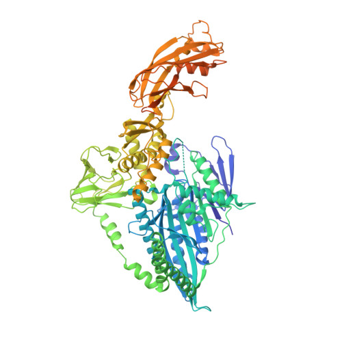
    7WRG

  • PubMed Abstract: 

    Autoinhibition of kinesin-3 ensures the proper spatiotemporal control of the motor activity for intracellular transport, but the underlying mechanism remains elusive. Here, we determine the full-length structure of kinesin-3 KLP-6 in a compact self-folded state. Unexpectedly, all the internal coiled-coil segments and domains in KLP-6 cooperate to successively lock down the neck and motor domains. The first coiled-coil segment is melted into several short helices that work with the motor domain to restrain the entire neck domain. The second coiled-coil segment associates with its neighboring FHA and MBS domains and integrates with the tail MATH domain to form a supramodule that synergistically wraps around the motor domain to trap the nucleotide and hinder the microtubule binding. This multilevel-lockdown mechanism for autoinhibition could be applicable to other kinesin-3 motors.


  • Organizational Affiliation
    • National Laboratory of Biomacromolecules, CAS Center for Excellence in Biomacromolecules, Institute of Biophysics, Chinese Academy of Sciences, 15 Datun Road, 100101, Beijing, China.

Macromolecule Content 

  • Total Structure Weight: 215.55 kDa 
  • Atom Count: 12,589 
  • Modeled Residue Count: 1,595 
  • Deposited Residue Count: 1,920 
  • Unique protein chains: 1

Macromolecules

Find similar proteins by:|  3D Structure
Entity ID: 1
MoleculeChains  Sequence LengthOrganismDetailsImage
Kinesin-like protein
A, B
960Caenorhabditis elegansMutation(s): 0 
Gene Names: klp-6CELE_R144.1R144.1
UniProt
Find proteins for G5EFQ4 (Caenorhabditis elegans)
Explore G5EFQ4 
Go to UniProtKB:  G5EFQ4
Entity Groups
Sequence Clusters30% Identity50% Identity70% Identity90% Identity95% Identity100% Identity
UniProt GroupG5EFQ4
Sequence Annotations
Expand
Reference Sequence

Experimental Data & Validation

Experimental Data

  • Method: X-RAY DIFFRACTION
  • Resolution: 3.16 Å
  • R-Value Free:  0.263 (Depositor), 0.259 (DCC) 
  • R-Value Work:  0.213 (Depositor), 0.214 (DCC) 
  • R-Value Observed: 0.216 (Depositor) 
Space Group: P 1 21 1
Unit Cell:
Length ( Å )Angle ( ˚ )
a = 56.138α = 90
b = 68.816β = 92.75
c = 253.216γ = 90
Software Package:
Software NamePurpose
PHENIXrefinement
PDB_EXTRACTdata extraction
HKL-2000data reduction
HKL-2000data scaling
PHASERphasing

Structure Validation

View Full Validation Report


Ligand Structure Quality Assessment 


Entry History 

& Funding Information

Deposition Data


Funding OrganizationLocationGrant Number
Chinese Academy of SciencesChina--

Revision History  (Full details and data files)

  • Version 1.0: 2022-08-10
    Type: Initial release
  • Version 1.1: 2023-11-29
    Changes: Data collection, Refinement description