7WKU | pdb_00007wku

Structure of PDCoV Mpro in complex with an inhibitor


Experimental Data Snapshot

  • Method: X-RAY DIFFRACTION
  • Resolution: 2.60 Å
  • R-Value Free: 
    0.241 (Depositor), 0.241 (DCC) 
  • R-Value Work: 
    0.191 (Depositor), 0.194 (DCC) 
  • R-Value Observed: 
    0.193 (Depositor) 

Starting Model: experimental
View more details

wwPDB Validation 3D Report Full Report

Validation slider image for 7WKU

This is version 2.2 of the entry. See complete history

Literature

The Structure of the Porcine Deltacoronavirus Main Protease Reveals a Conserved Target for the Design of Antivirals.

Wang, F.Chen, C.Wang, Z.Han, X.Shi, P.Zhou, K.Liu, X.Xiao, Y.Cai, Y.Huang, J.Zhang, L.Yang, H.

(2022) Viruses 14

  • DOI: https://doi.org/10.3390/v14030486
  • Primary Citation Related Structures: 
    7WKU

  • PubMed Abstract: 

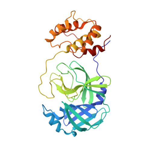
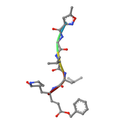
    The existing zoonotic coronaviruses (CoVs) and viral genetic variants are important microbiological pathogens that cause severe disease in humans and animals. Currently, no effective broad-spectrum antiviral drugs against existing and emerging CoVs are available. The CoV main protease (M pro ) plays an essential role in viral replication, making it an ideal target for drug development. However, the structure of the Deltacoronavirus M pro is still unavailable. Porcine deltacoronavirus (PDCoV) is a novel CoV that belongs to the genus Deltacoronavirus and causes atrophic enteritis, severe diarrhea, vomiting and dehydration in pigs. Here, we determined the structure of PDCoV M pro complexed with a Michael acceptor inhibitor. Structural comparison showed that the backbone of PDCoV M pro is similar to those of alpha-, beta- and gamma-CoV M pro s. The substrate-binding pocket of M pro is well conserved in the subfamily Coronavirinae . In addition, we also observed that M pro s from the same genus adopted a similar conformation. Furthermore, the structure of PDCoV M pro in complex with a Michael acceptor inhibitor revealed the mechanism of its inhibition of PDCoV M pro . Our results provide a basis for the development of broad-spectrum antivirals against PDCoV and other CoVs.


  • Organizational Affiliation
    • School of Life Sciences, Tianjin University, Tianjin 300072, China.

Macromolecule Content 

  • Total Structure Weight: 212.1 kDa 
  • Atom Count: 14,877 
  • Modeled Residue Count: 1,834 
  • Deposited Residue Count: 1,884 
  • Unique protein chains: 2

Macromolecules

Find similar proteins by:|  3D Structure
Entity ID: 1
MoleculeChains  Sequence LengthOrganismDetailsImage
Peptidase C30
A, B, C, D, E
A, B, C, D, E, F
308Porcine deltacoronavirusMutation(s): 0 
Gene Names: ORF1ab
UniProt
Find proteins for A0A140ESF0 (Porcine deltacoronavirus)
Explore A0A140ESF0 
Go to UniProtKB:  A0A140ESF0
Entity Groups
Sequence Clusters30% Identity50% Identity70% Identity90% Identity95% Identity100% Identity
UniProt GroupA0A140ESF0
Sequence Annotations
Expand
Reference Sequence
Find similar proteins by:  Sequence   |   3D Structure  
Entity ID: 2
MoleculeChains  Sequence LengthOrganismDetailsImage
N-[(5-METHYLISOXAZOL-3-YL)CARBONYL]ALANYL-L-VALYL-N~1~-((1R,2Z)-4-(BENZYLOXY)-4-OXO-1-{[(3R)-2-OXOPYRROLIDIN-3-YL]METHYL}BUT-2-ENYL)-L-LEUCINAMIDE6synthetic constructMutation(s): 0 

Experimental Data & Validation

Experimental Data

  • Method: X-RAY DIFFRACTION
  • Resolution: 2.60 Å
  • R-Value Free:  0.241 (Depositor), 0.241 (DCC) 
  • R-Value Work:  0.191 (Depositor), 0.194 (DCC) 
  • R-Value Observed: 0.193 (Depositor) 
Space Group: P 61
Unit Cell:
Length ( Å )Angle ( ˚ )
a = 122.292α = 90
b = 122.292β = 90
c = 289.754γ = 120
Software Package:
Software NamePurpose
PHENIXrefinement
HKL-2000data scaling
PDB_EXTRACTdata extraction
PHENIXmodel building
PHENIXphasing

Structure Validation

View Full Validation Report


Entry History 

& Funding Information

Deposition Data


Funding OrganizationLocationGrant Number
National Natural Science Foundation of China (NSFC)China81772204

Revision History  (Full details and data files)

  • Version 1.0: 2022-05-25
    Type: Initial release
  • Version 2.0: 2023-11-15
    Changes: Atomic model, Data collection, Derived calculations
  • Version 2.1: 2023-11-29
    Changes: Refinement description
  • Version 2.2: 2024-10-16
    Changes: Structure summary