7WDT | pdb_00007wdt

6-sulfo-beta-D-N-acetylglucosaminidase from Bifidobacterium bifidum in complex with GlcNAc-6S


Experimental Data Snapshot

  • Method: X-RAY DIFFRACTION
  • Resolution: 1.65 Å
  • R-Value Free: 
    0.200 (Depositor), 0.212 (DCC) 
  • R-Value Work: 
    0.170 (Depositor), 0.184 (DCC) 
  • R-Value Observed: 
    0.171 (Depositor) 

wwPDB Validation 3D Report Full Report

Validation slider image for 7WDT

Ligand Structure Quality Assessment 


This is version 1.3 of the entry. See complete history

Literature

A bacterial sulfoglycosidase highlights mucin O-glycan breakdown in the gut ecosystem.

Katoh, T.Yamada, C.Wallace, M.D.Yoshida, A.Gotoh, A.Arai, M.Maeshibu, T.Kashima, T.Hagenbeek, A.Ojima, M.N.Takada, H.Sakanaka, M.Shimizu, H.Nishiyama, K.Ashida, H.Hirose, J.Suarez-Diez, M.Nishiyama, M.Kimura, I.Stubbs, K.A.Fushinobu, S.Katayama, T.

(2023) Nat Chem Biol 19: 778-789

  • DOI: https://doi.org/10.1038/s41589-023-01272-y
  • Primary Citation Related Structures: 
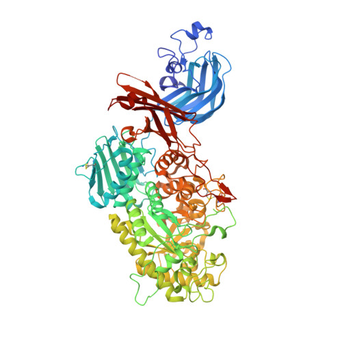
    7WDT, 7WDU

  • PubMed Abstract: 

    Mucinolytic bacteria modulate host-microbiota symbiosis and dysbiosis through their ability to degrade mucin O-glycans. However, how and to what extent bacterial enzymes are involved in the breakdown process remains poorly understood. Here we focus on a glycoside hydrolase family 20 sulfoglycosidase (BbhII) from Bifidobacterium bifidum, which releases N-acetylglucosamine-6-sulfate from sulfated mucins. Glycomic analysis showed that, in addition to sulfatases, sulfoglycosidases are involved in mucin O-glycan breakdown in vivo and that the released N-acetylglucosamine-6-sulfate potentially affects gut microbial metabolism, both of which were also supported by a metagenomic data mining analysis. Enzymatic and structural analysis of BbhII reveals the architecture underlying its specificity and the presence of a GlcNAc-6S-specific carbohydrate-binding module (CBM) 32 with a distinct sugar recognition mode that B. bifidum takes advantage of to degrade mucin O-glycans. Comparative analysis of the genomes of prominent mucinolytic bacteria also highlights a CBM-dependent O-glycan breakdown strategy used by B. bifidum.


  • Organizational Affiliation
    • Graduate School of Biostudies, Kyoto University, Sakyo-ku, Kyoto, Japan.

Macromolecule Content 

  • Total Structure Weight: 90.42 kDa 
  • Atom Count: 6,897 
  • Modeled Residue Count: 819 
  • Deposited Residue Count: 830 
  • Unique protein chains: 1

Macromolecules

Find similar proteins by:|  3D Structure
Entity ID: 1
MoleculeChains  Sequence LengthOrganismDetailsImage
Beta-N-acetylhexosaminidase830Bifidobacterium bifidum JCM 1254Mutation(s): 0 
Gene Names: bbhII
EC: 3.2.1.52
UniProt
Find proteins for D4QAP5 (Bifidobacterium bifidum)
Explore D4QAP5 
Go to UniProtKB:  D4QAP5
Entity Groups
Sequence Clusters30% Identity50% Identity70% Identity90% Identity95% Identity100% Identity
UniProt GroupD4QAP5
Sequence Annotations
Expand
Reference Sequence

Experimental Data & Validation

Experimental Data

  • Method: X-RAY DIFFRACTION
  • Resolution: 1.65 Å
  • R-Value Free:  0.200 (Depositor), 0.212 (DCC) 
  • R-Value Work:  0.170 (Depositor), 0.184 (DCC) 
  • R-Value Observed: 0.171 (Depositor) 
Space Group: P 1 21 1
Unit Cell:
Length ( Å )Angle ( ˚ )
a = 78.131α = 90
b = 57.879β = 98.59
c = 98.53γ = 90
Software Package:
Software NamePurpose
REFMACrefinement
PDB_EXTRACTdata extraction
XDSdata reduction
Aimlessdata scaling
PHENIXphasing

Structure Validation

View Full Validation Report


Ligand Structure Quality Assessment 


Entry History 

& Funding Information

Deposition Data


Funding OrganizationLocationGrant Number
Japan Society for the Promotion of Science (JSPS)Japan19K05789
Japan Society for the Promotion of Science (JSPS)Japan21H02116

Revision History  (Full details and data files)

  • Version 1.0: 2022-12-28
    Type: Initial release
  • Version 1.1: 2023-03-15
    Changes: Database references
  • Version 1.2: 2023-06-14
    Changes: Database references
  • Version 1.3: 2024-11-13
    Changes: Data collection, Structure summary