7W8L | pdb_00007w8l

Crystal Structure of Co-type nitrile hydratase mutant from Pseudonocardia thermophila - M46R


Experimental Data Snapshot

  • Method: X-RAY DIFFRACTION
  • Resolution: 2.30 Å
  • R-Value Free: 
    0.233 (Depositor), 0.195 (DCC) 
  • R-Value Work: 
    0.180 (Depositor), 0.186 (DCC) 
  • R-Value Observed: 
    0.182 (Depositor) 

Starting Model: experimental
View more details

wwPDB Validation 3D Report Full Report

Validation slider image for 7W8L

This is version 1.2 of the entry. See complete history

Literature

Insight into the broadened substrate scope of nitrile hydratase by static and dynamic structure analysis.

Ma, D.Cheng, Z.Peplowski, L.Han, L.Xia, Y.Hou, X.Guo, J.Yin, D.Rao, Y.Zhou, Z.

(2022) Chem Sci 13: 8417-8428

  • DOI: https://doi.org/10.1039/d2sc02319a
  • Primary Citation Related Structures: 
    7W8L, 7W8M

  • PubMed Abstract: 

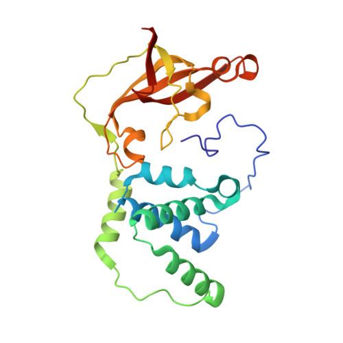
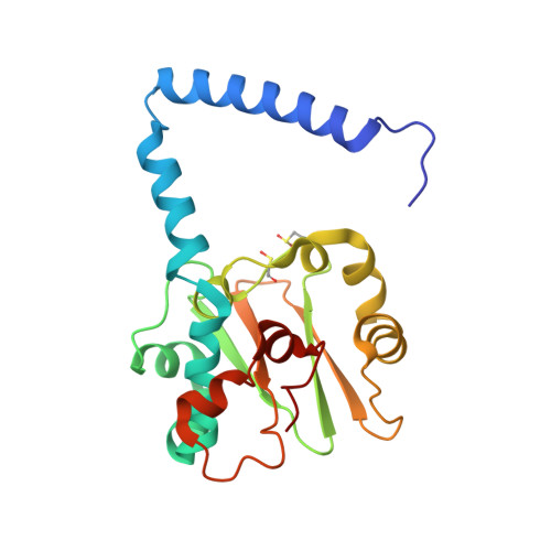
    The narrow substrate scope limits the wide industrial application of enzymes. Here, we successfully broadened the substrate scope of a nitrile hydratase (NHase) through mutation of two tunnel entrance residues based on rational tunnel calculation. Two variants, with increased specific activity, especially toward bulky substrates, were obtained. Crystal structure analysis revealed that the mutations led to the expansion of the tunnel entrance, which might be conducive to substrate entry. More importantly, molecular dynamics simulations illustrated that the mutations introduced anti-correlated movements to the regions around the substrate tunnel and the active site, which would promote substrate access during the dynamic process of catalysis. Additionally, mutations on the corresponding tunnel entrance residues on other NHases also enhanced their activity toward bulky substrates. These results not only revealed that residues located at the enzyme surface were a key factor in enzyme catalytic performance, but also provided dynamic evidence for insight into enzyme substrate scope broadening.


  • Organizational Affiliation
    • Key Laboratory of Industrial Biotechnology (Ministry of Education), School of Biotechnology, Jiangnan University Wuxi Jiangsu 214122 China raoyijian@jiangnan.edu.cn zhmzhou@jiangnan.edu.cn.

Macromolecule Content 

  • Total Structure Weight: 49.96 kDa 
  • Atom Count: 3,651 
  • Modeled Residue Count: 428 
  • Deposited Residue Count: 438 
  • Unique protein chains: 2

Macromolecules

Find similar proteins by:|  3D Structure
Entity ID: 1
MoleculeChains  Sequence LengthOrganismDetailsImage
Cobalt-containing nitrile hydratase subunit betaA [auth B]233Pseudonocardia thermophila DSM 43832Mutation(s): 1 
EC: 4.2.1.84
UniProt
Find proteins for Q7SID3 (Pseudonocardia thermophila)
Explore Q7SID3 
Go to UniProtKB:  Q7SID3
Entity Groups
Sequence Clusters30% Identity50% Identity70% Identity90% Identity95% Identity100% Identity
UniProt GroupQ7SID3
Sequence Annotations
Expand
Reference Sequence
Find similar proteins by:|  3D Structure
Entity ID: 2
MoleculeChains  Sequence LengthOrganismDetailsImage
Nitrile hydrataseB [auth A]205Pseudonocardia thermophila DSM 43832Mutation(s): 0 
Gene Names: SAMN05443637_10361
EC: 4.2.1.84
UniProt
Find proteins for Q7SID2 (Pseudonocardia thermophila)
Explore Q7SID2 
Go to UniProtKB:  Q7SID2
Entity Groups
Sequence Clusters30% Identity50% Identity70% Identity90% Identity95% Identity100% Identity
UniProt GroupQ7SID2
Sequence Annotations
Expand
Reference Sequence

Small Molecules

Ligands 1 Unique
IDChains Name / Formula / InChI Key2D Diagram3D Interactions
CO
(Subject of Investigation/LOI)

Query on CO


Download:Ideal Coordinates CCD File
C [auth A]COBALT (II) ION
Co
XLJKHNWPARRRJB-UHFFFAOYSA-N
Modified Residues  2 Unique
IDChains TypeFormula2D DiagramParent
CSD
Query on CSD
B [auth A]L-PEPTIDE LINKINGC3 H7 N O4 SCYS
CSO
Query on CSO
B [auth A]L-PEPTIDE LINKINGC3 H7 N O3 SCYS

Experimental Data & Validation

Experimental Data

  • Method: X-RAY DIFFRACTION
  • Resolution: 2.30 Å
  • R-Value Free:  0.233 (Depositor), 0.195 (DCC) 
  • R-Value Work:  0.180 (Depositor), 0.186 (DCC) 
  • R-Value Observed: 0.182 (Depositor) 
Space Group: P 32 2 1
Unit Cell:
Length ( Å )Angle ( ˚ )
a = 65.461α = 90
b = 65.461β = 90
c = 182.805γ = 120
Software Package:
Software NamePurpose
PHENIXrefinement
SAINTdata scaling
PDB_EXTRACTdata extraction
SAINTdata reduction
PHENIXphasing

Structure Validation

View Full Validation Report


Entry History 

& Funding Information

Deposition Data


Funding OrganizationLocationGrant Number
National Natural Science Foundation of China (NSFC)China--

Revision History  (Full details and data files)

  • Version 1.0: 2022-08-24
    Type: Initial release
  • Version 1.1: 2023-11-29
    Changes: Data collection, Refinement description
  • Version 1.2: 2024-11-06
    Changes: Structure summary