7VW6 | pdb_00007vw6

Cryo-EM Structure of Formate Dehydrogenase 1 from Methylorubrum extorquens AM1


Experimental Data Snapshot

  • Method: ELECTRON MICROSCOPY
  • Resolution: 2.19 Å
  • Aggregation State: PARTICLE 
  • Reconstruction Method: SINGLE PARTICLE 

wwPDB Validation 3D Report Full Report

Validation slider image for 7VW6

This is version 1.2 of the entry. See complete history

Literature

Multiple electron transfer pathways of tungsten-containing formate dehydrogenase in direct electron transfer-type bioelectrocatalysis.

Yoshikawa, T.Makino, F.Miyata, T.Suzuki, Y.Tanaka, H.Namba, K.Kano, K.Sowa, K.Kitazumi, Y.Shirai, O.

(2022) Chem Commun (Camb) 58: 6478-6481

  • DOI: https://doi.org/10.1039/d2cc01541b
  • Primary Citation Related Structures: 
    7VW6

  • PubMed Abstract: 

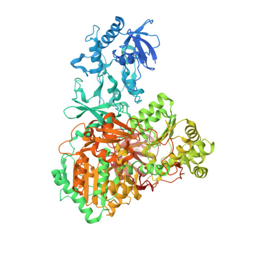
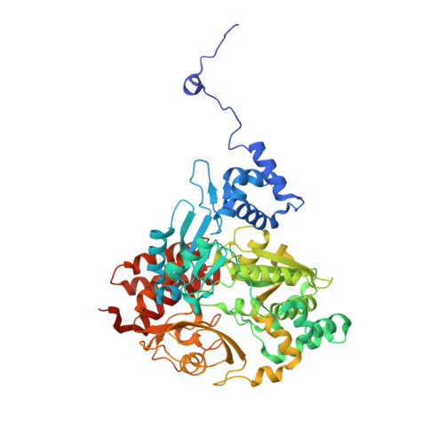
    Tungsten-containing formate dehydrogenase from Methylorubrum extroquens AM1 (FoDH1)-a promising biocatalyst for the interconversion of carbon dioxide/formate and nicotine adenine dinucleotide (NAD + )/NADH redox couples-was investigated using structural biology and bioelectrochemistry. FoDH1 is reported to be an enzyme that can realize "direct electron transfer (DET)-type bioelectrocatalysis." However, its 3-D structure, electrode-active sites, and electron transfer (ET) pathways remain unclear. The ET pathways were investigated using structural information, electrostatic interactions between the electrode and the enzyme, and the differences in the substrates. Two electrode-active sites and multiple ET pathways in FoDH1 were discovered.


  • Organizational Affiliation
    • Division of Applied Life Sciences, Graduate School of Agriculture, Kyoto University, Sakyo Kyoto, 606-8502, Japan. sowa.keisei.2u@kyoto-u.ac.jp.

Macromolecule Content 

  • Total Structure Weight: 173.74 kDa 
  • Atom Count: 11,485 
  • Modeled Residue Count: 1,473 
  • Deposited Residue Count: 1,561 
  • Unique protein chains: 2

Macromolecules

Find similar proteins by:|  3D Structure
Entity ID: 1
MoleculeChains  Sequence LengthOrganismDetailsImage
Formate dehydrogenase989Methylorubrum extorquens AM1Mutation(s): 0 
EC: 1.17.1.9 (PDB Primary Data), 1.2.1.2 (UniProt)
UniProt
Find proteins for C5ATT7 (Methylorubrum extorquens (strain ATCC 14718 / DSM 1338 / JCM 2805 / NCIMB 9133 / AM1))
Explore C5ATT7 
Go to UniProtKB:  C5ATT7
Entity Groups
Sequence Clusters30% Identity50% Identity70% Identity90% Identity95% Identity100% Identity
UniProt GroupC5ATT7
Sequence Annotations
Expand
Reference Sequence
Find similar proteins by:|  3D Structure
Entity ID: 2
MoleculeChains  Sequence LengthOrganismDetailsImage
Tungsten-containing formate dehydrogenase beta subunit572Methylorubrum extorquens AM1Mutation(s): 0 
EC: 1.2.1.2
UniProt
Find proteins for C5ATT6 (Methylorubrum extorquens (strain ATCC 14718 / DSM 1338 / JCM 2805 / NCIMB 9133 / AM1))
Explore C5ATT6 
Go to UniProtKB:  C5ATT6
Entity Groups
Sequence Clusters30% Identity50% Identity70% Identity90% Identity95% Identity100% Identity
UniProt GroupC5ATT6
Sequence Annotations
Expand
Reference Sequence

Small Molecules

Ligands 6 Unique
IDChains Name / Formula / InChI Key2D Diagram3D Interactions
MGD

Query on MGD


Download:Ideal Coordinates CCD File
G [auth A],
H [auth A]
2-AMINO-5,6-DIMERCAPTO-7-METHYL-3,7,8A,9-TETRAHYDRO-8-OXA-1,3,9,10-TETRAAZA-ANTHRACEN-4-ONE GUANOSINE DINUCLEOTIDE
C20 H26 N10 O13 P2 S2
VQAGYJCYOLHZDH-ILXWUORBSA-N
FMN
(Subject of Investigation/LOI)

Query on FMN


Download:Ideal Coordinates CCD File
M [auth B]FLAVIN MONONUCLEOTIDE
C17 H21 N4 O9 P
FVTCRASFADXXNN-SCRDCRAPSA-N
SF4

Query on SF4


Download:Ideal Coordinates CCD File
C [auth A],
D [auth A],
F [auth A],
L [auth B]
IRON/SULFUR CLUSTER
Fe4 S4
LJBDFODJNLIPKO-UHFFFAOYSA-N
W

Query on W


Download:Ideal Coordinates CCD File
I [auth A]TUNGSTEN ION
W
FZFRVZDLZISPFJ-UHFFFAOYSA-N
FES

Query on FES


Download:Ideal Coordinates CCD File
E [auth A],
K [auth B]
FE2/S2 (INORGANIC) CLUSTER
Fe2 S2
NIXDOXVAJZFRNF-UHFFFAOYSA-N
UNX

Query on UNX


Download:Ideal Coordinates CCD File
J [auth A]UNKNOWN ATOM OR ION
X

Experimental Data & Validation

Experimental Data

  • Method: ELECTRON MICROSCOPY
  • Resolution: 2.19 Å
  • Aggregation State: PARTICLE 
  • Reconstruction Method: SINGLE PARTICLE 
EM Software:
TaskSoftware PackageVersion
RECONSTRUCTIONRELION3.1
MODEL REFINEMENTPHENIX1.18

Structure Validation

View Full Validation Report


Entry History 

& Funding Information

Deposition Data


Funding OrganizationLocationGrant Number
Japan Agency for Medical Research and Development (AMED)JapanJP21am0101117
Japan Society for the Promotion of Science (JSPS)JapanJP21H01961

Revision History  (Full details and data files)

  • Version 1.0: 2022-06-01
    Type: Initial release
  • Version 1.1: 2022-06-15
    Changes: Database references
  • Version 1.2: 2024-06-26
    Changes: Data collection