7VRU

Crystal structure of PacII_M1M2S-DNA-SAH complex


Experimental Data Snapshot

  • Method: X-RAY DIFFRACTION
  • Resolution: 2.40 Å
  • R-Value Free: 0.235 
  • R-Value Work: 0.190 
  • R-Value Observed: 0.192 

wwPDB Validation   3D Report Full Report


Ligand Structure Quality Assessment 


This is version 1.0 of the entry. See complete history


Literature

Molecular insights into DNA recognition and methylation by non-canonical type I restriction-modification systems.

Zhu, J.Gao, Y.Wang, Y.Zhan, Q.Feng, H.Luo, X.Li, P.Liu, S.Hou, H.Gao, P.

(2022) Nat Commun 13: 6391-6391

  • DOI: https://doi.org/10.1038/s41467-022-34085-z
  • Primary Citation of Related Structures:  
    7VRU, 7VS4

  • PubMed Abstract: 

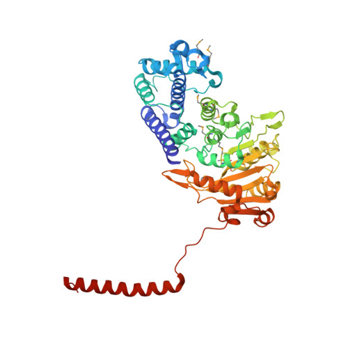
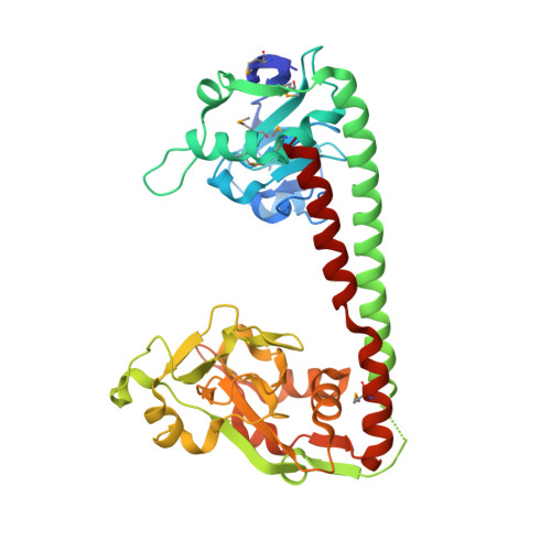
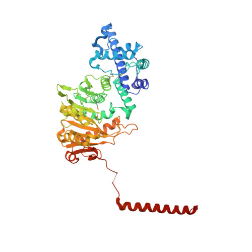
    Type I restriction-modification systems help establish the prokaryotic DNA methylation landscape and provide protection against invasive DNA. In addition to classical m6A modifications, non-canonical type I enzymes catalyze both m6A and m4C using alternative DNA-modification subunits M1 and M2. Here, we report the crystal structures of the non-canonical PacII_M1M2S methyltransferase bound to target DNA and reaction product S-adenosylhomocysteine in a closed clamp-like conformation. Target DNA binds tightly within the central tunnel of the M1M2S complex and forms extensive contacts with all three protein subunits. Unexpectedly, while the target cytosine properly inserts into M2's pocket, the target adenine (either unmethylated or methylated) is anchored outside M1's pocket. A unique asymmetric catalysis is established where PacII_M1M2S has precisely coordinated the relative conformations of different subunits and evolved specific amino acids within M2/M1. This work provides insights into mechanisms of m6A/m4C catalysis and guidance for designing tools based on type I restriction-modification enzymes.


  • Organizational Affiliation

    CAS Key Laboratory of Infection and Immunity, CAS Center for Excellence in Biomacromolecules, Institute of Biophysics, Chinese Academy of Sciences, Beijing, 100101, China.


Macromolecules

Find similar proteins by:  (by identity cutoff)  |  3D Structure
Entity ID: 1
MoleculeChains Sequence LengthOrganismDetailsImage
Site-specific DNA-methyltransferase (adenine-specific)A [auth B]504Pseudomonas alcaligenesMutation(s): 0 
Gene Names: A0T30_13470
Entity Groups  
Sequence Clusters30% Identity50% Identity70% Identity90% Identity95% Identity100% Identity
Sequence Annotations
Expand
  • Reference Sequence
Find similar proteins by:  (by identity cutoff)  |  3D Structure
Entity ID: 2
MoleculeChains Sequence LengthOrganismDetailsImage
Site-specific DNA recognition subunitB [auth C]383Pseudomonas alcaligenesMutation(s): 0 
Gene Names: pacIIS
Entity Groups  
Sequence Clusters30% Identity50% Identity70% Identity90% Identity95% Identity100% Identity
Sequence Annotations
Expand
  • Reference Sequence
Find similar proteins by:  (by identity cutoff)  |  3D Structure
Entity ID: 5
MoleculeChains Sequence LengthOrganismDetailsImage
Site-specific DNA-methyltransferase (adenine-specific)E [auth A]499Pseudomonas alcaligenesMutation(s): 0 
Gene Names: A0T30_13480
EC: 2.1.1.72
Entity Groups  
Sequence Clusters30% Identity50% Identity70% Identity90% Identity95% Identity100% Identity
Sequence Annotations
Expand
  • Reference Sequence

Find similar nucleic acids by:  Sequence   |   3D Structure  

Entity ID: 3
MoleculeChains LengthOrganismImage
DNA (25-mer)C [auth H]25synthetic construct
Sequence Annotations
Expand
  • Reference Sequence

Find similar nucleic acids by:  Sequence   |   3D Structure  

Entity ID: 4
MoleculeChains LengthOrganismImage
DNA (25-mer)D [auth I]25synthetic construct
Sequence Annotations
Expand
  • Reference Sequence
Small Molecules
Modified Residues  1 Unique
IDChains TypeFormula2D DiagramParent
MSE
Query on MSE
A [auth B]L-PEPTIDE LINKINGC5 H11 N O2 SeMET
Experimental Data & Validation

Experimental Data

  • Method: X-RAY DIFFRACTION
  • Resolution: 2.40 Å
  • R-Value Free: 0.235 
  • R-Value Work: 0.190 
  • R-Value Observed: 0.192 
  • Space Group: P 21 21 21
Unit Cell:
Length ( Å )Angle ( ˚ )
a = 109.8α = 90
b = 122.655β = 90
c = 131.361γ = 90
Software Package:
Software NamePurpose
PHENIXrefinement
HKL-2000data reduction
HKL-2000data scaling
PHENIXphasing
Cootmodel building

Structure Validation

View Full Validation Report


Ligand Structure Quality Assessment 


Entry History & Funding Information

Deposition Data


Funding OrganizationLocationGrant Number
National Natural Science Foundation of China (NSFC)China31922037

Revision History  (Full details and data files)

  • Version 1.0: 2022-11-30
    Type: Initial release