7VOS | pdb_00007vos

High-resolution neutron and X-ray joint refined structure of high-potential iron-sulfur protein in the oxidized state


Experimental Data Snapshot

  • Method: X-RAY DIFFRACTION
  • Resolution: 0.66 Å
  • R-Value Free: 
    0.082 (Depositor) 
  • R-Value Work: 
    0.074 (Depositor) 
  • R-Value Observed: 
    0.074 (Depositor) 

  • Method: NEUTRON DIFFRACTION
  • Resolution: 1.20 Å
  • R-Value Free: 
    0.168 (Depositor) 
  • R-Value Work: 
    0.154 (Depositor) 
  • R-Value Observed: 
    0.154 (Depositor) 

Starting Model: experimental
View more details

wwPDB Validation 3D Report Full Report

Validation slider image for 7VOS

This is version 1.1 of the entry. See complete history

Literature

Revisiting the concept of peptide bond planarity in an iron-sulfur protein by neutron structure analysis.

Hanazono, Y.Hirano, Y.Takeda, K.Kusaka, K.Tamada, T.Miki, K.

(2022) Sci Adv 8: eabn2276-eabn2276

  • DOI: https://doi.org/10.1126/sciadv.abn2276
  • Primary Citation Related Structures: 
    7VOS

  • PubMed Abstract: 

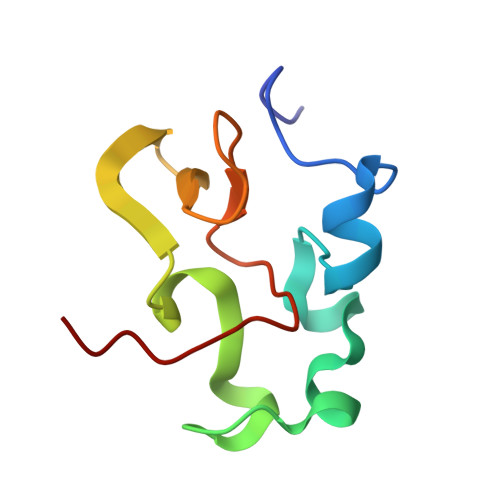
    The planarity of the peptide bond is important for the stability and structure formation of proteins. However, substantial distortion of peptide bonds has been reported in several high-resolution structures and computational analyses. To investigate the peptide bond planarity, including hydrogen atoms, we report a 1.2-Å resolution neutron structure of the oxidized form of high-potential iron-sulfur protein. This high-resolution neutron structure shows that the nucleus positions of the amide protons deviate from the peptide plane and shift toward the acceptors. The planarity of the H─N─C═O plane depends strongly on the pyramidalization of the nitrogen atom. Moreover, the orientation of the amide proton of Cys 75 is different in the reduced and oxidized states, possibly because of the electron storage capacity of the iron-sulfur cluster.


  • Organizational Affiliation
    • Department of Chemistry, Graduate School of Science, Kyoto University, Sakyo-ku, Kyoto 606-8502, Japan.

Macromolecule Content 

  • Total Structure Weight: 10.18 kDa 
  • Atom Count: 959 
  • Modeled Residue Count: 83 
  • Deposited Residue Count: 83 
  • Unique protein chains: 1

Macromolecules

Find similar proteins by:|  3D Structure
Entity ID: 1
MoleculeChains  Sequence LengthOrganismDetailsImage
High-potential iron-sulfur protein83Thermochromatium tepidumMutation(s): 0 
UniProt
Find proteins for P80176 (Thermochromatium tepidum)
Explore P80176 
Go to UniProtKB:  P80176
Entity Groups
Sequence Clusters30% Identity50% Identity70% Identity90% Identity95% Identity100% Identity
UniProt GroupP80176
Sequence Annotations
Expand
Reference Sequence

Small Molecules

Ligands 5 Unique
IDChains Name / Formula / InChI Key2D Diagram3D Interactions
SF4
(Subject of Investigation/LOI)

Query on SF4


Download:Ideal Coordinates CCD File
B [auth A]IRON/SULFUR CLUSTER
Fe4 S4
LJBDFODJNLIPKO-UHFFFAOYSA-N
SO4

Query on SO4


Download:Ideal Coordinates CCD File
C [auth A],
D [auth A],
E [auth A],
F [auth A],
G [auth A]
SULFATE ION
O4 S
QAOWNCQODCNURD-UHFFFAOYSA-L
GOL

Query on GOL


Download:Ideal Coordinates CCD File
H [auth A],
I [auth A],
J [auth A],
K [auth A],
L [auth A]
GLYCEROL
C3 H8 O3
PEDCQBHIVMGVHV-UHFFFAOYSA-N
D3O

Query on D3O


Download:Ideal Coordinates CCD File
Q [auth A]trideuteriooxidanium
D3 O
XLYOFNOQVPJJNP-ZRLBSURWSA-O
NH4

Query on NH4


Download:Ideal Coordinates CCD File
M [auth A],
N [auth A],
O [auth A],
P [auth A]
AMMONIUM ION
H4 N
QGZKDVFQNNGYKY-UHFFFAOYSA-O

Experimental Data & Validation

Experimental Data

  • Method: X-RAY DIFFRACTION
  • Resolution: 0.66 Å
  • R-Value Free:  0.082 (Depositor) 
  • R-Value Work:  0.074 (Depositor) 
  • R-Value Observed: 0.074 (Depositor) 
  • Method: NEUTRON DIFFRACTION
  • Resolution: 1.20 Å
  • R-Value Free:  0.168 (Depositor) 
  • R-Value Work:  0.154 (Depositor) 
  • R-Value Observed: 0.154 (Depositor) 
Space Group: P 21 21 21
Unit Cell:
Length ( Å )Angle ( ˚ )
a = 46.334α = 90
b = 58.855β = 90
c = 23.548γ = 90
Software Package:
Software NamePurpose
PHENIXrefinement
SCALAdata scaling
MOLREPphasing
HKL-2000data collection

Structure Validation

View Full Validation Report


Entry History 

& Funding Information

Deposition Data


Funding OrganizationLocationGrant Number
Ministry of Education, Culture, Sports, Science and Technology (Japan)Japan--
Japan Science and TechnologyJapan--
Japan Society for the Promotion of Science (JSPS)Japan--

Revision History  (Full details and data files)

  • Version 1.0: 2022-06-01
    Type: Initial release
  • Version 1.1: 2024-04-03
    Changes: Data collection, Refinement description