7VOK

The Crystal structure of EF-Tu and GDP from Mycobacterium tuberculosis


Experimental Data Snapshot

  • Method: X-RAY DIFFRACTION
  • Resolution: 3.40 Å
  • R-Value Free: 0.302 
  • R-Value Work: 0.244 
  • R-Value Observed: 0.247 

Starting Model: experimental
View more details

wwPDB Validation   3D Report Full Report


Ligand Structure Quality Assessment 


This is version 1.2 of the entry. See complete history


Literature

Structural insights of the elongation factor EF-Tu complexes in protein translation of Mycobacterium tuberculosis.

Zhan, B.Gao, Y.Gao, W.Li, Y.Li, Z.Qi, Q.Lan, X.Shen, H.Gan, J.Zhao, G.Li, J.

(2022) Commun Biol 5: 1052-1052

  • DOI: https://doi.org/10.1038/s42003-022-04019-y
  • Primary Citation of Related Structures:  
    7VMX, 7VOK

  • PubMed Abstract: 

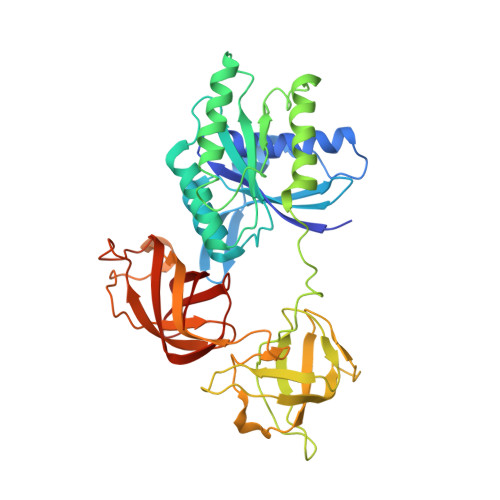
    Tuberculosis (TB) caused by Mycobacterium tuberculosis (Mtb) is the second-deadliest infectious disease worldwide. Emerging evidence shows that the elongation factor EF-Tu could be an excellent target for treating Mtb infection. Here, we report the crystal structures of Mtb EF-Tu•EF-Ts and EF-Tu•GDP complexes, showing the molecular basis of EF-Tu's representative recycling and inactive forms in protein translation. Mtb EF-Tu binds with EF-Ts at a 1:1 ratio in solution and crystal packing. Mutation and SAXS analysis show that EF-Ts residues Arg13, Asn82, and His149 are indispensable for the EF-Tu/EF-Ts complex formation. The GDP binding pocket of EF-Tu dramatically changes conformations upon binding with EF-Ts, sharing a similar GDP-exchange mechanism in E. coli and T. ther. Also, the FDA-approved drug Osimertinib inhibits the growth of M. smegmatis, H37Ra, and M. bovis BCG strains by directly binding with EF-Tu. Thus, our work reveals the structural basis of Mtb EF-Tu in polypeptide synthesis and may provide a promising candidate for TB treatment.


  • Organizational Affiliation

    State Key Laboratory of Genetic Engineering, School of Life Sciences, Shanghai Engineering Research Center of Industrial Microorganisms, Fudan University, 200438, Shanghai, China.


Macromolecules
Find similar proteins by:  (by identity cutoff)  |  3D Structure
Entity ID: 1
MoleculeChains Sequence LengthOrganismDetailsImage
Elongation factor Tu
A, B, C, D
396Mycobacterium tuberculosisMutation(s): 0 
Gene Names: 
UniProt
Find proteins for P9WNN1 (Mycobacterium tuberculosis (strain ATCC 25618 / H37Rv))
Explore P9WNN1 
Go to UniProtKB:  P9WNN1
Entity Groups  
Sequence Clusters30% Identity50% Identity70% Identity90% Identity95% Identity100% Identity
UniProt GroupP9WNN1
Sequence Annotations
Expand
  • Reference Sequence
Experimental Data & Validation

Experimental Data

  • Method: X-RAY DIFFRACTION
  • Resolution: 3.40 Å
  • R-Value Free: 0.302 
  • R-Value Work: 0.244 
  • R-Value Observed: 0.247 
  • Space Group: P 1 21 1
Unit Cell:
Length ( Å )Angle ( ˚ )
a = 95.059α = 90
b = 75.18β = 93.77
c = 127.176γ = 90
Software Package:
Software NamePurpose
PHENIXrefinement
HKL-3000data reduction
HKL-3000data scaling
PHASERphasing

Structure Validation

View Full Validation Report


Ligand Structure Quality Assessment 


Entry History & Funding Information

Deposition Data


Funding OrganizationLocationGrant Number
Ministry of Science and Technology (MoST, China)China2016YFA0500600

Revision History  (Full details and data files)

  • Version 1.0: 2022-10-19
    Type: Initial release
  • Version 1.1: 2023-11-29
    Changes: Data collection, Refinement description
  • Version 1.2: 2024-04-24
    Changes: Database references