


Conformational change of catalytic residue in reduced enzyme of FAD-dependent Glucose Dehydrogenase at pH6.5
Nakajima, Y.To be published.
Experimental Data Snapshot
Entity ID: 1 | |||||
|---|---|---|---|---|---|
| Molecule | Chains | Sequence Length | Organism | Details | Image |
| GMC oxidoreductase | 572 | Aspergillus oryzae | Mutation(s): 0 Gene Names: OAory_01010120 EC: 1.1.3.4 | 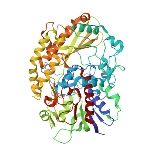 | |
UniProt | |||||
Entity Groups | |||||
| Sequence Clusters | 30% Identity50% Identity70% Identity90% Identity95% Identity100% Identity | ||||
| UniProt Group | A0A1S9DW10 | ||||
Sequence AnnotationsExpand | |||||
Reference Sequence | |||||
| Ligands 3 Unique | |||||
|---|---|---|---|---|---|
| ID | Chains | Name / Formula / InChI Key | 2D Diagram | 3D Interactions | |
| FDA (Subject of Investigation/LOI) Download:Ideal Coordinates CCD File | C [auth A] | DIHYDROFLAVINE-ADENINE DINUCLEOTIDE C27 H35 N9 O15 P2 YPZRHBJKEMOYQH-UYBVJOGSSA-N |  | ||
| B3P Download:Ideal Coordinates CCD File | D [auth A] | 2-[3-(2-HYDROXY-1,1-DIHYDROXYMETHYL-ETHYLAMINO)-PROPYLAMINO]-2-HYDROXYMETHYL-PROPANE-1,3-DIOL C11 H26 N2 O6 HHKZCCWKTZRCCL-UHFFFAOYSA-N |  | ||
| LGC (Subject of Investigation/LOI) Download:Ideal Coordinates CCD File | B [auth A] | D-glucono-1,5-lactone C6 H10 O6 PHOQVHQSTUBQQK-SQOUGZDYSA-N |  | ||
| Length ( Å ) | Angle ( ˚ ) |
|---|---|
| a = 95.421 | α = 90 |
| b = 95.421 | β = 90 |
| c = 123.841 | γ = 90 |
| Software Name | Purpose |
|---|---|
| REFMAC | refinement |
| HKL-2000 | data reduction |
| HKL-2000 | data scaling |
| MOLREP | phasing |
| Funding Organization | Location | Grant Number |
|---|---|---|
| Ministry of Education, Culture, Sports, Science and Technology (Japan) | Japan | 24780106 |


