7VBQ | pdb_00007vbq

Heterodimer structure of Fe(II)/(alpha)ketoglutarate-dependent dioxygenase TlxIJ


Experimental Data Snapshot

  • Method: X-RAY DIFFRACTION
  • Resolution: 1.95 Å
  • R-Value Free: 
    0.214 (Depositor), 0.210 (DCC) 
  • R-Value Work: 
    0.171 (Depositor), 0.169 (DCC) 
  • R-Value Observed: 
    0.173 (Depositor) 

Starting Model: experimental
View more details

wwPDB Validation 3D Report Full Report

Validation slider image for 7VBQ

Ligand Structure Quality Assessment 


This is version 1.1 of the entry. See complete history

Literature

Heterodimeric Non-heme Iron Enzymes in Fungal Meroterpenoid Biosynthesis.

Li, X.Awakawa, T.Mori, T.Ling, M.Hu, D.Wu, B.Abe, I.

(2021) J Am Chem Soc 143: 21425-21432

  • DOI: https://doi.org/10.1021/jacs.1c11548
  • Primary Citation Related Structures: 
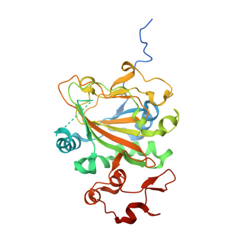
    7VBQ, 7VBR

  • PubMed Abstract: 

    Talaromyolides ( 1 - 6 ) are a group of unusual 6/6/6/6/6/6 hexacyclic meroterpenoids with (3 R )-6-hydroxymellein and 4,5-seco-drimane substructures, isolated from the marine fungus Talaromyces purpureogenus . We have identified the biosynthetic gene cluster tlxA-J by heterologous expression in Aspergillus , in vitro enzyme assays, and CRISPR-Cas9-based gene inactivation. Remarkably, the heterodimer of non-heme iron (NHI) enzymes, TlxJ-TlxI, catalyzes three steps of oxidation including a key reaction, hydroxylation at C-5 and C-9 of 12 , the intermediate with 3-ketohydroxydrimane scaffold, to facilitate a retro-aldol reaction, leading to the construction of the 4,5-secodrimane skeleton and characteristic ketal scaffold of 1 - 6 . The products of TlxJ-TlxI, 1 and 4 , were further hydroxylated at C-4'β by another NHI heterodimer, TlxA-TlxC, and acetylated by TlxB to yield the final products, 3 and 6 . The X-ray structural analysis coupled with site-directed mutagenesis provided insights into the heterodimer TlxJ-TlxI formation and its catalysis. This is the first report to show that two NHI proteins form a heterodimer for catalysis and utilizes a novel methodology to create functional oxygenase structures in secondary metabolite biosynthesis.


  • Organizational Affiliation
    • Graduate School of Pharmaceutical Sciences, The University of Tokyo, Bunkyo-ku, Tokyo 113-0033, Japan.

Macromolecule Content 

  • Total Structure Weight: 67.9 kDa 
  • Atom Count: 4,773 
  • Modeled Residue Count: 547 
  • Deposited Residue Count: 600 
  • Unique protein chains: 2

Macromolecules

Find similar proteins by:|  3D Structure
Entity ID: 1
MoleculeChains  Sequence LengthOrganismDetailsImage
Fe(II)/(alpha)ketoglutarate-dependent dioxygenase TlxJ318Talaromyces purpureogenusMutation(s): 0 
Find similar proteins by:|  3D Structure
Entity ID: 2
MoleculeChains  Sequence LengthOrganismDetailsImage
Fe(II)/(alpha)ketoglutarate-dependent dioxygenase TlxI282Talaromyces purpureogenusMutation(s): 0 

Experimental Data & Validation

Experimental Data

  • Method: X-RAY DIFFRACTION
  • Resolution: 1.95 Å
  • R-Value Free:  0.214 (Depositor), 0.210 (DCC) 
  • R-Value Work:  0.171 (Depositor), 0.169 (DCC) 
  • R-Value Observed: 0.173 (Depositor) 
Space Group: P 21 21 21
Unit Cell:
Length ( Å )Angle ( ˚ )
a = 58.933α = 90
b = 75.335β = 90
c = 133.611γ = 90
Software Package:
Software NamePurpose
PHENIXrefinement
XDSdata reduction
Aimlessdata scaling
PHASERphasing

Structure Validation

View Full Validation Report


Ligand Structure Quality Assessment 


Entry History 

& Funding Information

Deposition Data


Funding OrganizationLocationGrant Number
Not funded--

Revision History  (Full details and data files)

  • Version 1.0: 2022-04-20
    Type: Initial release
  • Version 1.1: 2023-11-29
    Changes: Data collection, Refinement description