


Enzymatic catalysis favours eight-membered over five-membered ring closure in bicyclomycin biosynthesis
He, J.B., Wu, L., Wei, W., Meng, S., Liu, Z.T., Wu, X., Pan, H.X., Yang, S., Liang, Y., Zhou, J., Tang, G.L.(2023) Nat Catal 6: 637-648
Experimental Data Snapshot
Starting Model: experimental
View more details
Entity ID: 1 | |||||
|---|---|---|---|---|---|
| Molecule | Chains | Sequence Length | Organism | Details | Image |
| dioxygenase | 328 | Streptomyces ossamyceticus | Mutation(s): 0 | 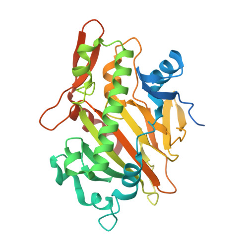 | |
UniProt | |||||
Find proteins for A0A9Y2YA87 (Streptomyces ossamyceticus) Explore A0A9Y2YA87 Go to UniProtKB: A0A9Y2YA87 | |||||
Entity Groups | |||||
| Sequence Clusters | 30% Identity50% Identity70% Identity90% Identity95% Identity100% Identity | ||||
| UniProt Group | A0A9Y2YA87 | ||||
Sequence AnnotationsExpand | |||||
Reference Sequence | |||||
| Ligands 4 Unique | |||||
|---|---|---|---|---|---|
| ID | Chains | Name / Formula / InChI Key | 2D Diagram | 3D Interactions | |
| 7QX (Subject of Investigation/LOI) Download:Ideal Coordinates CCD File | E [auth A] | (3S,6S)-3-[(2S)-butan-2-yl]-6-[(2R)-2-methyl-2,3-bis(oxidanyl)propyl]piperazine-2,5-dion C12 H22 N2 O4 KJJLKZILTAEYLN-PHGLEFOZSA-N |  | ||
| AKG (Subject of Investigation/LOI) Download:Ideal Coordinates CCD File | B [auth A] | 2-OXOGLUTARIC ACID C5 H6 O5 KPGXRSRHYNQIFN-UHFFFAOYSA-N |  | ||
| GOL Download:Ideal Coordinates CCD File | D [auth A] | GLYCEROL C3 H8 O3 PEDCQBHIVMGVHV-UHFFFAOYSA-N |  | ||
| FE2 Download:Ideal Coordinates CCD File | C [auth A] | FE (II) ION Fe CWYNVVGOOAEACU-UHFFFAOYSA-N |  | ||
| Length ( Å ) | Angle ( ˚ ) |
|---|---|
| a = 101.097 | α = 90 |
| b = 101.097 | β = 90 |
| c = 130.377 | γ = 120 |
| Software Name | Purpose |
|---|---|
| PHENIX | refinement |
| XDS | data reduction |
| pointless | data scaling |
| PHASER | phasing |
| Funding Organization | Location | Grant Number |
|---|---|---|
| Not funded | -- |


