7UWR | pdb_00007uwr

KSQ+AT from first module of the pikromycin synthase


Experimental Data Snapshot

  • Method: ELECTRON MICROSCOPY
  • Resolution: 2.61 Å
  • Aggregation State: PARTICLE 
  • Reconstruction Method: SINGLE PARTICLE 

Starting Model: experimental
View more details

wwPDB Validation 3D Report Full Report

Validation slider image for 7UWR

This is version 1.3 of the entry. See complete history

Literature

Priming enzymes from the pikromycin synthase reveal how assembly-line ketosynthases catalyze carbon-carbon chemistry.

Dickinson, M.S.Miyazawa, T.McCool, R.S.Keatinge-Clay, A.T.

(2022) Structure 30: 1331

  • DOI: https://doi.org/10.1016/j.str.2022.05.021
  • Primary Citation Related Structures: 
    7UWR, 8CZC

  • PubMed Abstract: 

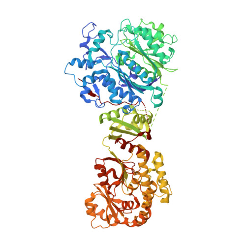
    The first domain of modular polyketide synthases (PKSs) is most commonly a ketosynthase (KS)-like enzyme, KS Q , that primes polyketide synthesis. Unlike downstream KSs that fuse α-carboxyacyl groups to growing polyketide chains, it performs an extension-decoupled decarboxylation of these groups to generate primer units. When Pik127, a model triketide synthase constructed from modules of the pikromycin synthase, was studied by cryoelectron microscopy (cryo-EM), the dimeric didomain comprised of KS Q and the neighboring methylmalonyl-selective acyltransferase (AT) dominated the class averages and yielded structures at 2.5- and 2.8-Å resolution, respectively. Comparisons with ketosynthases complexed with their substrates revealed the conformation of the (2S)-methylmalonyl-S-phosphopantetheinyl portion of KS Q and KS substrates prior to decarboxylation. Point mutants of Pik127 probed the roles of residues in the KS Q active site, while an AT-swapped version of Pik127 demonstrated that KS Q can also decarboxylate malonyl groups. Mechanisms for how KS Q and KS domains catalyze carbon-carbon chemistry are proposed.


  • Organizational Affiliation
    • Sauer Structural Biology Lab, The University of Texas at Austin, 102 E. 24th Street, Austin, TX 78712, USA.

Macromolecule Content 

  • Total Structure Weight: 183.6 kDa 
  • Atom Count: 12,361 
  • Modeled Residue Count: 1,674 
  • Deposited Residue Count: 1,772 
  • Unique protein chains: 1

Macromolecules

Find similar proteins by:|  3D Structure
Entity ID: 1
MoleculeChains  Sequence LengthOrganismDetailsImage
Narbonolide/10-deoxymethynolide synthase PikA1, modules 1 and 2
A, B
886Streptomyces venezuelaeMutation(s): 0 
Gene Names: pikAI
EC: 2.3.1.239 (PDB Primary Data), 2.3.1.240 (PDB Primary Data)
UniProt
Find proteins for Q9ZGI5 (Streptomyces venezuelae)
Explore Q9ZGI5 
Go to UniProtKB:  Q9ZGI5
Entity Groups
Sequence Clusters30% Identity50% Identity70% Identity90% Identity95% Identity100% Identity
UniProt GroupQ9ZGI5
Sequence Annotations
Expand
Reference Sequence

Experimental Data & Validation

Experimental Data

  • Method: ELECTRON MICROSCOPY
  • Resolution: 2.61 Å
  • Aggregation State: PARTICLE 
  • Reconstruction Method: SINGLE PARTICLE 
EM Software:
TaskSoftware PackageVersion
RECONSTRUCTIONcryoSPARC3.2.0
MODEL REFINEMENTCoot0.8.9.2
MODEL REFINEMENTPHENIX1.19.2-4158

Structure Validation

View Full Validation Report


Entry History 

& Funding Information

Deposition Data


Funding OrganizationLocationGrant Number
National Institutes of Health/National Institute of General Medical Sciences (NIH/NIGMS)United StatesGM106112
Welch FoundationUnited StatesF-1712

Revision History  (Full details and data files)

  • Version 1.0: 2022-06-08
    Type: Initial release
  • Version 1.1: 2022-07-06
    Changes: Database references
  • Version 1.2: 2022-09-14
    Changes: Database references
  • Version 1.3: 2024-06-12
    Changes: Data collection, Refinement description