7URX | pdb_00007urx

SJ25C1 Fab in complex with soluble CD19


Experimental Data Snapshot

  • Method: ELECTRON MICROSCOPY
  • Resolution: 3.40 Å
  • Aggregation State: PARTICLE 
  • Reconstruction Method: SINGLE PARTICLE 

wwPDB Validation 3D Report Full Report

Validation slider image for 7URX

This is version 1.3 of the entry. See complete history

Literature

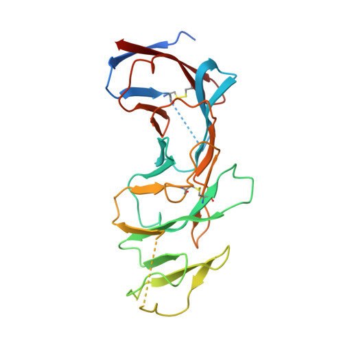
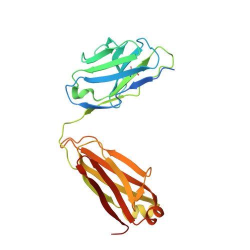
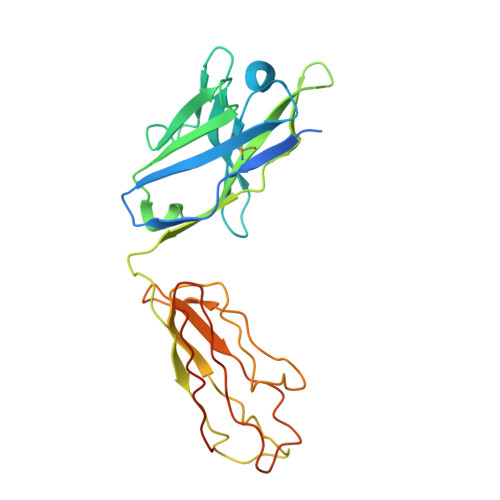
CD19 CAR antigen engagement mechanisms and affinity tuning.

He, C.Mansilla-Soto, J.Khanra, N.Hamieh, M.Bustos, V.Paquette, A.J.Garcia Angus, A.Shore, D.M.Rice, W.J.Khelashvili, G.Sadelain, M.Meyerson, J.R.

(2023) Sci Immunol 8: eadf1426-eadf1426

  • DOI: https://doi.org/10.1126/sciimmunol.adf1426
  • Primary Citation Related Structures: 
    7URV, 7URX

  • PubMed Abstract: 

    Chimeric antigen receptor (CAR) T cell therapy relies on T cells that are guided by synthetic receptors to target and lyse cancer cells. CARs bind to cell surface antigens through an scFv (binder), the affinity of which is central to determining CAR T cell function and therapeutic success. CAR T cells targeting CD19 were the first to achieve marked clinical responses in patients with relapsed/refractory B cell malignancies and to be approved by the U.S. Food and Drug Administration (FDA). We report cryo-EM structures of CD19 antigen with the binder FMC63, which is used in four FDA-approved CAR T cell therapies (Kymriah, Yescarta, Tecartus, and Breyanzi), and the binder SJ25C1, which has also been used extensively in multiple clinical trials. We used these structures for molecular dynamics simulations, which guided creation of lower- or higher-affinity binders, and ultimately produced CAR T cells endowed with distinct tumor recognition sensitivities. The CAR T cells exhibited different antigen density requirements to trigger cytolysis and differed in their propensity to prompt trogocytosis upon contacting tumor cells. Our work shows how structural information can be applied to tune CAR T cell performance to specific target antigen densities.


  • Organizational Affiliation
    • Department of Physiology and Biophysics, Weill Cornell Medical College, New York, NY, USA.

Macromolecule Content 

  • Total Structure Weight: 83.45 kDa 
  • Atom Count: 4,492 
  • Modeled Residue Count: 655 
  • Deposited Residue Count: 765 
  • Unique protein chains: 3

Macromolecules

Find similar proteins by:|  3D Structure
Entity ID: 1
MoleculeChains  Sequence LengthOrganismDetailsImage
B-lymphocyte antigen CD19A [auth C]278Homo sapiensMutation(s): 0 
Gene Names: CD19
UniProt & NIH Common Fund Data Resources
Find proteins for P15391 (Homo sapiens)
Explore P15391 
Go to UniProtKB:  P15391
PHAROS:  P15391
GTEx:  ENSG00000177455 
Entity Groups
Sequence Clusters30% Identity50% Identity70% Identity90% Identity95% Identity100% Identity
UniProt GroupP15391
Sequence Annotations
Expand
Reference Sequence
Find similar proteins by:|  3D Structure
Entity ID: 2
MoleculeChains  Sequence LengthOrganismDetailsImage
SJ25C1 Fab light chainB [auth L]235Mus musculusMutation(s): 0 
Find similar proteins by:|  3D Structure
Entity ID: 3
MoleculeChains  Sequence LengthOrganismDetailsImage
SJ25C1 Fab heavy chainC [auth H]252Mus musculusMutation(s): 0 

Experimental Data & Validation

Experimental Data

  • Method: ELECTRON MICROSCOPY
  • Resolution: 3.40 Å
  • Aggregation State: PARTICLE 
  • Reconstruction Method: SINGLE PARTICLE 
EM Software:
TaskSoftware PackageVersion
MODEL REFINEMENTPHENIX

Structure Validation

View Full Validation Report


Entry History 

& Funding Information

Deposition Data


Funding OrganizationLocationGrant Number
Not funded--

Revision History  (Full details and data files)

  • Version 1.0: 2023-02-15
    Type: Initial release
  • Version 1.1: 2023-03-15
    Changes: Database references
  • Version 1.2: 2024-11-20
    Changes: Data collection, Structure summary
  • Version 1.3: 2025-05-21
    Changes: Data collection