7UFA

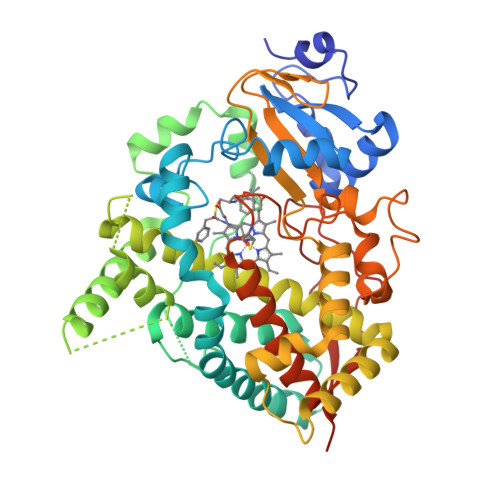
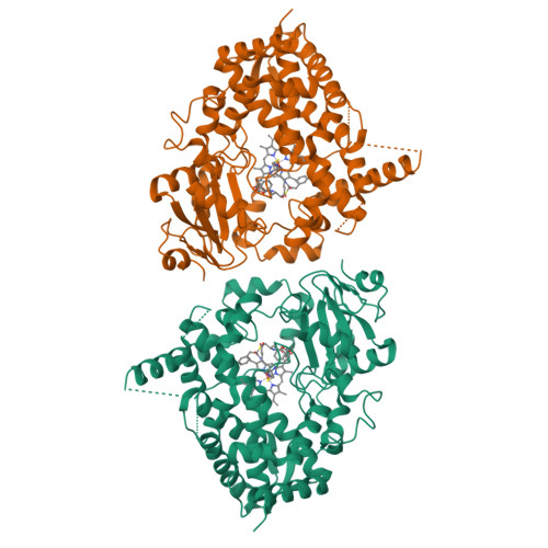
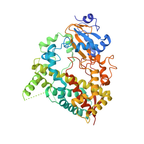
CYP3A4 bound to an inhibitor


Experimental Data Snapshot

  • Method: X-RAY DIFFRACTION
  • Resolution: 2.50 Å
  • R-Value Free: 0.283 
  • R-Value Work: 0.247 
  • R-Value Observed: 0.249 

Starting Model: experimental
View more details

wwPDB Validation   3D Report Full Report


Ligand Structure Quality Assessment 


This is version 1.1 of the entry. See complete history


Literature

Interaction of CYP3A4 with Rationally Designed Ritonavir Analogues: Impact of Steric Constraints Imposed on the Heme-Ligating Group and the End-Pyridine Attachment.

Samuels, E.R.Sevrioukova, I.F.

(2022) Int J Mol Sci 23

  • DOI: https://doi.org/10.3390/ijms23137291
  • Primary Citation of Related Structures:  
    7UF9, 7UFA, 7UFB, 7UFC, 7UFD, 7UFE, 7UFF

  • PubMed Abstract: 

    Controlled inhibition of drug-metabolizing cytochrome P450 3A4 (CYP3A4) is utilized to boost bioavailability of anti-viral and immunosuppressant pharmaceuticals. We investigate structure-activity relationships (SARs) in analogues of ritonavir, a potent CYP3A4 inhibitor marketed as pharmacoenhancer, to determine structural elements required for potent inhibition and whether the inhibitory potency can be further improved via a rational structure-based design. This study investigated eight (series VI) inhibitors differing in head- and end-moieties and their respective linkers. SAR analysis revealed the multifactorial regulation of inhibitory strength, with steric constraints imposed on the tethered heme-ligating moiety being a key factor. Minimization of these constraints by changing the linkers' length/flexibility and N-heteroatom position strengthened heme coordination and markedly improved binding and/or inhibitory strength. Impact of the end-pyridine attachment was not uniform due to influence of other determinants controlling the ligand-binding mode. This interplay between pharmacophoric determinants and the end-group enlargement can be used for further inhibitor optimization.


  • Organizational Affiliation

    Department of Pharmaceutical Sciences, University of California, Irvine, CA 92697, USA.


Macromolecules
Find similar proteins by:  (by identity cutoff)  |  3D Structure
Entity ID: 1
MoleculeChains Sequence LengthOrganismDetailsImage
Cytochrome P450 3A4487Homo sapiensMutation(s): 0 
Gene Names: CYP3A4CYP3A3
EC: 1.14.14.1 (PDB Primary Data), 1.14.14.56 (PDB Primary Data), 1.14.14.73 (PDB Primary Data), 1.14.14.55 (PDB Primary Data)
UniProt & NIH Common Fund Data Resources
Find proteins for P08684 (Homo sapiens)
Go to UniProtKB:  P08684
PHAROS:  P08684
GTEx:  ENSG00000160868 
Sequence Annotations
Expand
  • Reference Sequence
Small Molecules
Ligands 2 Unique
IDChains Name / Formula / InChI Key2D Diagram3D Interactions
HEM
Query on HEM

Download Ideal Coordinates CCD File 
B [auth A]PROTOPORPHYRIN IX CONTAINING FE
C34 H32 Fe N4 O4
KABFMIBPWCXCRK-RGGAHWMASA-L
NI6 (Subject of Investigation/LOI)
Query on NI6

Download Ideal Coordinates CCD File 
C [auth A]tert-butyl [(2S)-1-{[(2R)-1-oxo-3-phenyl-1-{[3-(pyridin-4-yl)propyl]amino}propan-2-yl]sulfanyl}-3-phenylpropan-2-yl]carbamate
C31 H39 N3 O3 S
HIUHMRVLQNHDQV-WUFINQPMSA-N
Experimental Data & Validation

Experimental Data

  • Method: X-RAY DIFFRACTION
  • Resolution: 2.50 Å
  • R-Value Free: 0.283 
  • R-Value Work: 0.247 
  • R-Value Observed: 0.249 
  • Space Group: I 2 2 2
Unit Cell:
Length ( Å )Angle ( ˚ )
a = 76.58α = 90
b = 101.73β = 90
c = 125.51γ = 90
Software Package:
Software NamePurpose
PHENIXrefinement
iMOSFLMdata reduction
SCALAdata scaling
PHASERphasing

Structure Validation

View Full Validation Report


Ligand Structure Quality Assessment 


Entry History & Funding Information

Deposition Data


Funding OrganizationLocationGrant Number
National Institutes of Health/National Institute of Environmental Health Sciences (NIH/NIEHS)United StatesES025767

Revision History  (Full details and data files)

  • Version 1.0: 2022-07-27
    Type: Initial release
  • Version 1.1: 2023-10-18
    Changes: Data collection, Refinement description