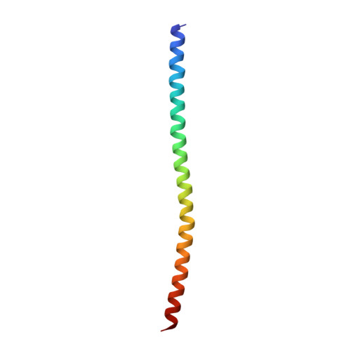
7UCD

Transcription factor FosB/JunD bZIP domain covalently modified with the cysteine-targeting alpha-haloketone compound Z2159931480


Experimental Data Snapshot

  • Method: X-RAY DIFFRACTION
  • Resolution: 3.21 Å
  • R-Value Free: 0.330 
  • R-Value Work: 0.280 
  • R-Value Observed: 0.284 

Starting Model: experimental
View more details

wwPDB Validation   3D Report Full Report


Ligand Structure Quality Assessment 


This is version 1.2 of the entry. See complete history


Literature

Chemically targeting the redox switch in AP1 transcription factor Delta FOSB.

Kumar, A.Aglyamova, G.Yim, Y.Y.Bailey, A.O.Lynch, H.M.Powell, R.T.Nguyen, N.D.Rosenthal, Z.Zhao, W.N.Li, Y.Chen, J.Fan, S.Lee, H.Russell, W.K.Stephan, C.Robison, A.J.Haggarty, S.J.Nestler, E.J.Zhou, J.Machius, M.Rudenko, G.

(2022) Nucleic Acids Res 50: 9548-9567

  • DOI: https://doi.org/10.1093/nar/gkac710
  • Primary Citation of Related Structures:  
    7UCC, 7UCD

  • PubMed Abstract: 

    The AP1 transcription factor ΔFOSB, a splice variant of FOSB, accumulates in the brain in response to chronic insults such as exposure to drugs of abuse, depression, Alzheimer's disease and tardive dyskinesias, and mediates subsequent long-term neuroadaptations. ΔFOSB forms heterodimers with other AP1 transcription factors, e.g. JUND, that bind DNA under control of a putative cysteine-based redox switch. Here, we reveal the structural basis of the redox switch by determining a key missing crystal structure in a trio, the ΔFOSB/JUND bZIP domains in the reduced, DNA-free form. Screening a cysteine-focused library containing 3200 thiol-reactive compounds, we identify specific compounds that target the redox switch, validate their activity biochemically and in cell-based assays, and show that they are well tolerated in different cell lines despite their general potential to bind to cysteines covalently. A crystal structure of the ΔFOSB/JUND bZIP domains in complex with a redox-switch-targeting compound reveals a deep compound-binding pocket near the DNA-binding site. We demonstrate that ΔFOSB, and potentially other, related AP1 transcription factors, can be targeted specifically and discriminately by exploiting unique structural features such as the redox switch and the binding partner to modulate biological function despite these proteins previously being thought to be undruggable.


  • Organizational Affiliation

    Department of Pharmacology and Toxicology, University of Texas Medical Branch, Galveston, TX 77555, USA.


Macromolecules
Find similar proteins by:  (by identity cutoff)  |  3D Structure
Entity ID: 1
MoleculeChains Sequence LengthOrganismDetailsImage
Protein fosBA [auth F]68Homo sapiensMutation(s): 0 
Gene Names: FOSBG0S3
UniProt & NIH Common Fund Data Resources
Find proteins for P53539 (Homo sapiens)
Explore P53539 
Go to UniProtKB:  P53539
PHAROS:  P53539
GTEx:  ENSG00000125740 
Entity Groups  
Sequence Clusters30% Identity50% Identity70% Identity90% Identity95% Identity100% Identity
UniProt GroupP53539
Sequence Annotations
Expand
  • Reference Sequence
Find similar proteins by:  (by identity cutoff)  |  3D Structure
Entity ID: 2
MoleculeChains Sequence LengthOrganismDetailsImage
Transcription factor jun-DB [auth J]68Homo sapiensMutation(s): 0 
Gene Names: JUND
UniProt & NIH Common Fund Data Resources
Find proteins for P17535 (Homo sapiens)
Explore P17535 
Go to UniProtKB:  P17535
PHAROS:  P17535
GTEx:  ENSG00000130522 
Entity Groups  
Sequence Clusters30% Identity50% Identity70% Identity90% Identity95% Identity100% Identity
UniProt GroupP17535
Sequence Annotations
Expand
  • Reference Sequence
Experimental Data & Validation

Experimental Data

  • Method: X-RAY DIFFRACTION
  • Resolution: 3.21 Å
  • R-Value Free: 0.330 
  • R-Value Work: 0.280 
  • R-Value Observed: 0.284 
  • Space Group: I 2 2 2
Unit Cell:
Length ( Å )Angle ( ˚ )
a = 48.26α = 90
b = 67.67β = 90
c = 122.943γ = 90
Software Package:
Software NamePurpose
PHENIXrefinement
XDSdata reduction
STARANISOdata scaling
PDB_EXTRACTdata extraction
PHASERphasing
Cootmodel building

Structure Validation

View Full Validation Report


Ligand Structure Quality Assessment 


Entry History & Funding Information

Deposition Data


Funding OrganizationLocationGrant Number
National Institutes of Health/National Institute on Drug Abuse (NIH/NIDA)United StatesR01DA040621

Revision History  (Full details and data files)

  • Version 1.0: 2023-01-25
    Type: Initial release
  • Version 1.1: 2023-10-25
    Changes: Data collection, Refinement description
  • Version 1.2: 2024-10-30
    Changes: Structure summary