


Structure of G6PD-WT dimer
Wei, X., Marmorstein, R.To be published.
Experimental Data Snapshot
wwPDB Validation 3D Report Full Report

Entity ID: 1 | |||||
|---|---|---|---|---|---|
| Molecule | Chains | Sequence Length | Organism | Details | Image |
| Glucose-6-phosphate 1-dehydrogenase | 523 | Homo sapiens | Mutation(s): 0 Gene Names: G6PD EC: 1.1.1.49 (PDB Primary Data), 1 (UniProt) | 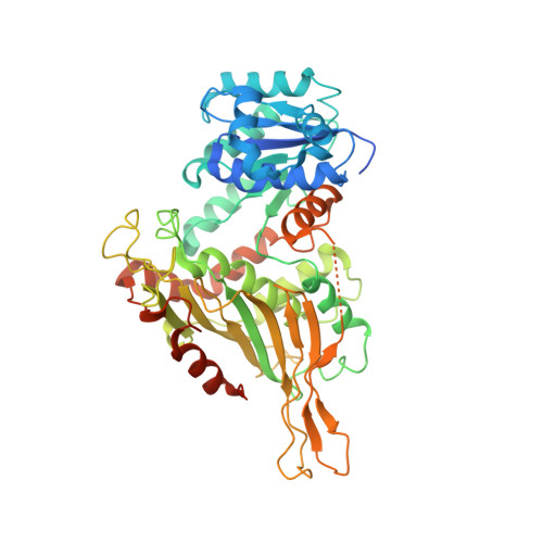 | |
UniProt & NIH Common Fund Data Resources | |||||
PHAROS: P11413 GTEx: ENSG00000160211 | |||||
Entity Groups | |||||
| Sequence Clusters | 30% Identity50% Identity70% Identity90% Identity95% Identity100% Identity | ||||
| UniProt Group | P11413 | ||||
Sequence AnnotationsExpand | |||||
Reference Sequence | |||||
| Task | Software Package | Version |
|---|---|---|
| MODEL REFINEMENT | PHENIX | |
| Funding Organization | Location | Grant Number |
|---|---|---|
| National Institutes of Health/National Institute of General Medical Sciences (NIH/NIGMS) | United States | -- |


