7U58 | pdb_00007u58

YcaO-mediated ATP-dependent peptidase activity in ribosomal peptide biosynthesis


Experimental Data Snapshot

  • Method: ELECTRON MICROSCOPY
  • Resolution: 3.10 Å
  • Aggregation State: 2D ARRAY 
  • Reconstruction Method: SINGLE PARTICLE 

wwPDB Validation 3D Report Full Report

Validation slider image for 7U58

This is version 1.2 of the entry. See complete history

Literature

YcaO-mediated ATP-dependent peptidase activity in ribosomal peptide biosynthesis.

Zheng, Y.Nair, S.K.

(2023) Nat Chem Biol 19: 111-119

  • DOI: https://doi.org/10.1038/s41589-022-01141-0
  • Primary Citation Related Structures: 
    7U58

  • PubMed Abstract: 

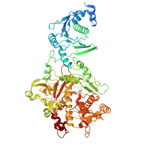
    YcaO enzymes catalyze ATP-dependent post-translation modifications on peptides, including the installation of (ox/thi)azoline, thioamide and/or amidine moieties. Here we demonstrate that, in the biosynthesis of the bis-methyloxazolic alkaloid muscoride A, the YcaO enzyme MusD carries out both ATP-dependent cyclodehydration and peptide bond cleavage, which is a mechanism unprecedented for such a reaction. YcaO-catalyzed modifications are proposed to occur through a backbone O-phosphorylated intermediate, but this mechanism remains speculative. We report, to our knowedge, the first characterization of an acyl-phosphate species consistent with the proposed mechanism for backbone amide activation. The 3.1-Å-resolution cryogenic electron microscopy structure of MusD along with biochemical analysis allow identification of residues that enable peptide cleavage reaction. Bioinformatics analysis identifies other cyanobactin pathways that may deploy bifunctional YcaO enzymes. Our structural, mutational and mechanistic studies expand the scope of modifications catalyzed by YcaO proteins to include peptide hydrolysis and provide evidence for a unifying mechanism for the catalytically diverse outcomes.


  • Organizational Affiliation
    • Department of Biochemistry, University of Illinois at Urbana-Champaign, Roger Adams Laboratory, Urbana, IL, USA.

Macromolecule Content 

  • Total Structure Weight: 177.92 kDa 
  • Atom Count: 10,087 
  • Modeled Residue Count: 1,292 
  • Deposited Residue Count: 1,568 
  • Unique protein chains: 1

Macromolecules

Find similar proteins by:|  3D Structure
Entity ID: 1
MoleculeChains  Sequence LengthOrganismDetailsImage
MusD
A, B
784Desmonostoc sp. PCC 7906Mutation(s): 0 
Gene Names: musD
UniProt
Find proteins for A0A5Q0TWV7 (Desmonostoc sp. (strain PCC 7906))
Explore A0A5Q0TWV7 
Go to UniProtKB:  A0A5Q0TWV7
Entity Groups
Sequence Clusters30% Identity50% Identity70% Identity90% Identity95% Identity100% Identity
UniProt GroupA0A5Q0TWV7
Sequence Annotations
Expand
Reference Sequence

Experimental Data & Validation

Experimental Data

  • Method: ELECTRON MICROSCOPY
  • Resolution: 3.10 Å
  • Aggregation State: 2D ARRAY 
  • Reconstruction Method: SINGLE PARTICLE 

Structure Validation

View Full Validation Report


Entry History 

& Funding Information

Deposition Data


Funding OrganizationLocationGrant Number
National Institutes of Health/National Institute of General Medical Sciences (NIH/NIGMS)United StatesR01 GM079038

Revision History  (Full details and data files)

  • Version 1.0: 2022-11-02
    Type: Initial release
  • Version 1.1: 2023-01-11
    Changes: Database references
  • Version 1.2: 2024-06-12
    Changes: Data collection