7TXH | pdb_00007txh

Human MRas Q71R in complex with human Shoc2 LRR domain M173I and human PP1Ca


Experimental Data Snapshot

  • Method: X-RAY DIFFRACTION
  • Resolution: 1.95 Å
  • R-Value Free: 
    0.210 (Depositor), 0.212 (DCC) 
  • R-Value Work: 
    0.167 (Depositor), 0.171 (DCC) 
  • R-Value Observed: 
    0.169 (Depositor) 

Starting Models: experimental
View more details

wwPDB Validation 3D Report Full Report

Validation slider image for 7TXH

Ligand Structure Quality Assessment 


This is version 1.3 of the entry. See complete history

Literature

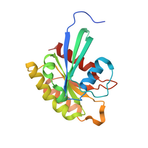
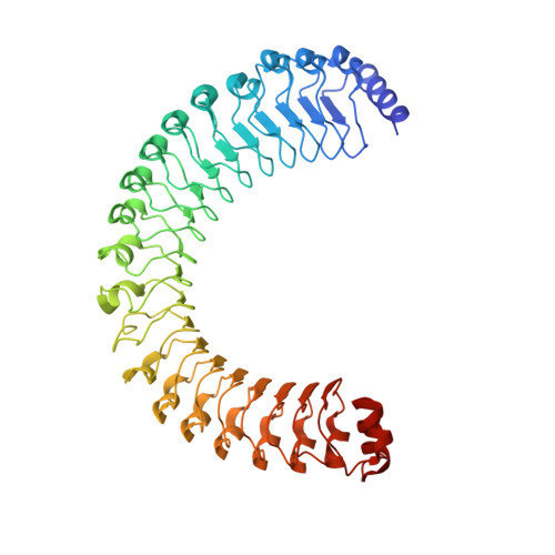
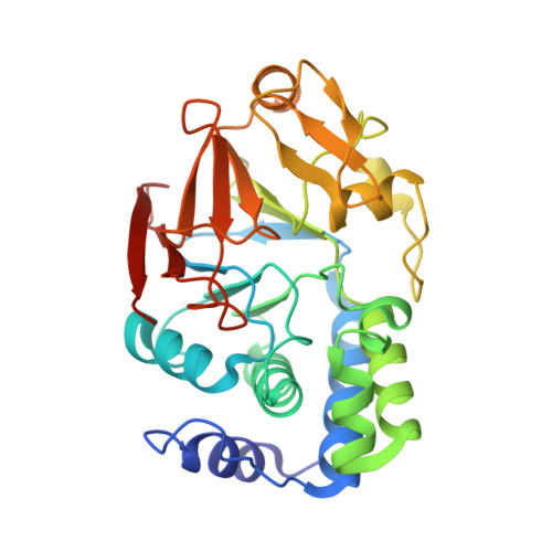
Structure of the MRAS-SHOC2-PP1C phosphatase complex.

Hauseman, Z.J.Fodor, M.Dhembi, A.Viscomi, J.Egli, D.Bleu, M.Katz, S.Park, E.Jang, D.M.Porter, K.A.Meili, F.Guo, H.Kerr, G.Molle, S.Velez-Vega, C.Beyer, K.S.Galli, G.G.Maira, S.M.Stams, T.Clark, K.Eck, M.J.Tordella, L.Thoma, C.R.King, D.A.

(2022) Nature 609: 416-423

  • DOI: https://doi.org/10.1038/s41586-022-05086-1
  • Primary Citation Related Structures: 
    7TXH, 7TYG

  • PubMed Abstract: 

    RAS-MAPK signalling is fundamental for cell proliferation and is altered in most human cancers 1-3 . However, our mechanistic understanding of how RAS signals through RAF is still incomplete. Although studies revealed snapshots for autoinhibited and active RAF-MEK1-14-3-3 complexes 4 , the intermediate steps that lead to RAF activation remain unclear. The MRAS-SHOC2-PP1C holophosphatase dephosphorylates RAF at serine 259, resulting in the partial displacement of 14-3-3 and RAF-RAS association 3,5,6 . MRAS, SHOC2 and PP1C are mutated in rasopathies-developmental syndromes caused by aberrant MAPK pathway activation 6-14 -and SHOC2 itself has emerged as potential target in receptor tyrosine kinase (RTK)-RAS-driven tumours 15-18 . Despite its importance, structural understanding of the SHOC2 holophosphatase is lacking. Here we determine, using X-ray crystallography, the structure of the MRAS-SHOC2-PP1C complex. SHOC2 bridges PP1C and MRAS through its concave surface and enables reciprocal interactions between all three subunits. Biophysical characterization indicates a cooperative assembly driven by the MRAS GTP-bound active state, an observation that is extendible to other RAS isoforms. Our findings support the concept of a RAS-driven and multi-molecular model for RAF activation in which individual RAS-GTP molecules recruit RAF-14-3-3 and SHOC2-PP1C to produce downstream pathway activation. Importantly, we find that rasopathy and cancer mutations reside at protein-protein interfaces within the holophosphatase, resulting in enhanced affinities and function. Collectively, our findings shed light on a fundamental mechanism of RAS biology and on mechanisms of clinically observed enhanced RAS-MAPK signalling, therefore providing the structural basis for therapeutic interventions.


  • Organizational Affiliation
    • Novartis Institutes for BioMedical Research, Cambridge, MA, USA. zachary.hauseman@novartis.com.

Macromolecule Content 

  • Total Structure Weight: 225.67 kDa 
  • Atom Count: 17,319 
  • Modeled Residue Count: 1,920 
  • Deposited Residue Count: 1,964 
  • Unique protein chains: 3

Macromolecules

Find similar proteins by:|  3D Structure
Entity ID: 1
MoleculeChains  Sequence LengthOrganismDetailsImage
Ras-related protein M-Ras
A, D
180Homo sapiensMutation(s): 1 
Gene Names: MRASRRAS3
EC: 3.6.5.2
UniProt & NIH Common Fund Data Resources
Find proteins for O14807 (Homo sapiens)
Explore O14807 
Go to UniProtKB:  O14807
PHAROS:  O14807
GTEx:  ENSG00000158186 
Entity Groups
Sequence Clusters30% Identity50% Identity70% Identity90% Identity95% Identity100% Identity
UniProt GroupO14807
Sequence Annotations
Expand
Reference Sequence
Find similar proteins by:|  3D Structure
Entity ID: 2
MoleculeChains  Sequence LengthOrganismDetailsImage
Leucine-rich repeat protein SHOC-2
B, E
505Homo sapiensMutation(s): 1 
Gene Names: SHOC2KIAA0862
UniProt & NIH Common Fund Data Resources
Find proteins for Q9UQ13 (Homo sapiens)
Explore Q9UQ13 
Go to UniProtKB:  Q9UQ13
PHAROS:  Q9UQ13
GTEx:  ENSG00000108061 
Entity Groups
Sequence Clusters30% Identity50% Identity70% Identity90% Identity95% Identity100% Identity
UniProt GroupQ9UQ13
Sequence Annotations
Expand
Reference Sequence
Find similar proteins by:|  3D Structure
Entity ID: 3
MoleculeChains  Sequence LengthOrganismDetailsImage
Serine/threonine-protein phosphatase PP1-alpha catalytic subunit
C, F
297Homo sapiensMutation(s): 0 
Gene Names: PPP1CAPPP1A
EC: 3.1.3.16
UniProt & NIH Common Fund Data Resources
Find proteins for P62136 (Homo sapiens)
Explore P62136 
Go to UniProtKB:  P62136
PHAROS:  P62136
GTEx:  ENSG00000172531 
Entity Groups
Sequence Clusters30% Identity50% Identity70% Identity90% Identity95% Identity100% Identity
UniProt GroupP62136
Sequence Annotations
Expand
Reference Sequence

Small Molecules

Ligands 5 Unique
IDChains Name / Formula / InChI Key2D Diagram3D Interactions
GNP

Query on GNP


Download:Ideal Coordinates CCD File
G [auth A],
S [auth D]
PHOSPHOAMINOPHOSPHONIC ACID-GUANYLATE ESTER
C10 H17 N6 O13 P3
UQABYHGXWYXDTK-UUOKFMHZSA-N
PO4

Query on PO4


Download:Ideal Coordinates CCD File
BA [auth E],
EA [auth F],
O [auth C],
R [auth C]
PHOSPHATE ION
O4 P
NBIIXXVUZAFLBC-UHFFFAOYSA-K
GOL

Query on GOL


Download:Ideal Coordinates CCD File
AA [auth E]
FA [auth F]
GA [auth F]
HA [auth F]
I [auth A]
AA [auth E],
FA [auth F],
GA [auth F],
HA [auth F],
I [auth A],
J [auth B],
K [auth B],
L [auth B],
P [auth C],
Q [auth C],
U [auth D],
V [auth D],
W [auth E],
X [auth E],
Y [auth E],
Z [auth E]
GLYCEROL
C3 H8 O3
PEDCQBHIVMGVHV-UHFFFAOYSA-N
MN

Query on MN


Download:Ideal Coordinates CCD File
CA [auth F],
DA [auth F],
M [auth C],
N [auth C]
MANGANESE (II) ION
Mn
WAEMQWOKJMHJLA-UHFFFAOYSA-N
MG

Query on MG


Download:Ideal Coordinates CCD File
H [auth A],
T [auth D]
MAGNESIUM ION
Mg
JLVVSXFLKOJNIY-UHFFFAOYSA-N

Experimental Data & Validation

Experimental Data

  • Method: X-RAY DIFFRACTION
  • Resolution: 1.95 Å
  • R-Value Free:  0.210 (Depositor), 0.212 (DCC) 
  • R-Value Work:  0.167 (Depositor), 0.171 (DCC) 
  • R-Value Observed: 0.169 (Depositor) 
Space Group: P 1 21 1
Unit Cell:
Length ( Å )Angle ( ˚ )
a = 72.959α = 90
b = 115.755β = 100.314
c = 137.804γ = 90
Software Package:
Software NamePurpose
BUSTERrefinement
PHENIXrefinement
PHASERphasing
autoPROCdata reduction
autoPROCdata scaling

Structure Validation

View Full Validation Report


Ligand Structure Quality Assessment 


Entry History 

& Funding Information

Deposition Data


Funding OrganizationLocationGrant Number
Not funded--

Revision History  (Full details and data files)

  • Version 1.0: 2022-06-22
    Type: Initial release
  • Version 1.1: 2022-08-17
    Changes: Database references
  • Version 1.2: 2022-09-21
    Changes: Database references
  • Version 1.3: 2023-10-18
    Changes: Data collection, Refinement description