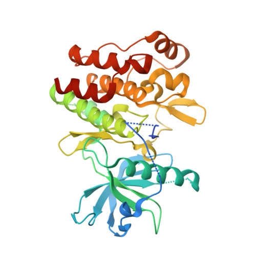
7TNH

Crystal structure of CSF1R kinase domain in complex with DP-6233


Experimental Data Snapshot

  • Method: X-RAY DIFFRACTION
  • Resolution: 2.70 Å
  • R-Value Free: 0.269 
  • R-Value Work: 0.215 
  • R-Value Observed: 0.217 

Starting Model: experimental
View more details

wwPDB Validation   3D Report Full Report


Ligand Structure Quality Assessment 


This is version 1.2 of the entry. See complete history


Literature

Discovery of acyl ureas as highly selective small molecule CSF1R kinase inhibitors.

Caldwell, T.M.Kaufman, M.D.Wise, S.C.Mi Ahn, Y.Hood, M.M.Lu, W.P.Patt, W.C.Samarakoon, T.Vogeti, L.Vogeti, S.Yates, K.M.Bulfer, S.L.Le Bourdonnec, B.Smith, B.D.Flynn, D.L.

(2022) Bioorg Med Chem Lett 74: 128929-128929

  • DOI: https://doi.org/10.1016/j.bmcl.2022.128929
  • Primary Citation of Related Structures:  
    7TNH

  • PubMed Abstract: 

    Based on the structure of an early lead identified in Deciphera's proprietary compound collection of switch control kinase inhibitors and using a combination of medicinal chemistry guided structure activity relationships and structure-based drug design, a novel series of potent acyl urea-based CSF1R inhibitors was identified displaying high selectivity for CSF1R versus the other members of the Type III receptor tyrosine kinase (RTK) family members (KIT, PDGFR-α, PDGFR-β, and FLT3), VEGFR2 and MET. Based on in vitro biology, in vitro ADME and in vivo PK/PD studies, compound 10 was selected as an advanced lead for Deciphera's CSF1R research program.


  • Organizational Affiliation

    Deciphera Pharmaceuticals, LLC, Waltham, MA 02451, United States.


Macromolecules
Find similar proteins by:  (by identity cutoff)  |  3D Structure
Entity ID: 1
MoleculeChains Sequence LengthOrganismDetailsImage
Macrophage colony-stimulating factor 1 receptor,Fibroblast growth factor receptor 1 chimera338Homo sapiensMutation(s): 0 
Gene Names: CSF1RFMSFGFR1BFGFRCEKFGFBRFLGFLT2HBGFR
EC: 2.7.10.1
UniProt & NIH Common Fund Data Resources
Find proteins for P11362 (Homo sapiens)
Explore P11362 
Go to UniProtKB:  P11362
PHAROS:  P11362
GTEx:  ENSG00000077782 
Find proteins for P07333 (Homo sapiens)
Explore P07333 
Go to UniProtKB:  P07333
PHAROS:  P07333
GTEx:  ENSG00000182578 
Entity Groups  
Sequence Clusters30% Identity50% Identity70% Identity90% Identity95% Identity100% Identity
UniProt GroupsP11362P07333
Sequence Annotations
Expand
  • Reference Sequence
Small Molecules
Ligands 3 Unique
IDChains Name / Formula / InChI Key2D Diagram3D Interactions
I9W (Subject of Investigation/LOI)
Query on I9W

Download Ideal Coordinates CCD File 
G [auth A]2,2-dimethyl-N-[(6-methyl-5-{[2-(1-methyl-1H-pyrazol-4-yl)pyridin-4-yl]oxy}pyridin-2-yl)carbamoyl]propanamide
C21 H24 N6 O3
WYLCQKJYTVNVCY-UHFFFAOYSA-N
CL
Query on CL

Download Ideal Coordinates CCD File 
C [auth A],
D [auth A],
E [auth A],
F [auth A]
CHLORIDE ION
Cl
VEXZGXHMUGYJMC-UHFFFAOYSA-M
NA
Query on NA

Download Ideal Coordinates CCD File 
B [auth A]SODIUM ION
Na
FKNQFGJONOIPTF-UHFFFAOYSA-N
Experimental Data & Validation

Experimental Data

  • Method: X-RAY DIFFRACTION
  • Resolution: 2.70 Å
  • R-Value Free: 0.269 
  • R-Value Work: 0.215 
  • R-Value Observed: 0.217 
  • Space Group: H 3
Unit Cell:
Length ( Å )Angle ( ˚ )
a = 81.08α = 90
b = 81.08β = 90
c = 146.67γ = 120
Software Package:
Software NamePurpose
XSCALEdata scaling
REFMACrefinement
PDB_EXTRACTdata extraction
XDSdata reduction
PHASERphasing

Structure Validation

View Full Validation Report


Ligand Structure Quality Assessment 


Entry History & Funding Information

Deposition Data


Funding OrganizationLocationGrant Number
Not funded--

Revision History  (Full details and data files)

  • Version 1.0: 2022-08-24
    Type: Initial release
  • Version 1.1: 2022-08-31
    Changes: Database references
  • Version 1.2: 2023-10-18
    Changes: Data collection, Refinement description