7TL7 | pdb_00007tl7

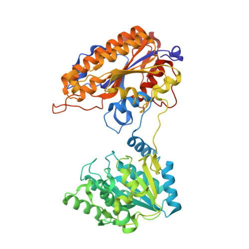
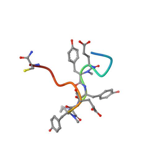
1.90A resolution structure of independent phosphoglycerate mutase from C. elegans in complex with a macrocyclic peptide inhibitor (Sa-D2)


Experimental Data Snapshot

  • Method: X-RAY DIFFRACTION
  • Resolution: 1.90 Å
  • R-Value Free: 
    0.216 (Depositor), 0.221 (DCC) 
  • R-Value Work: 
    0.171 (Depositor), 0.178 (DCC) 
  • R-Value Observed: 
    0.173 (Depositor) 

Starting Model: experimental
View more details

wwPDB Validation 3D Report Full Report

Validation slider image for 7TL7

This is version 1.2 of the entry. See complete history. 

Literature

Serum-Stable and Selective Backbone-N-Methylated Cyclic Peptides That Inhibit Prokaryotic Glycolytic Mutases.

van Neer, R.H.P., Dranchak, P.K., Liu, L., Aitha, M., Queme, B., Kimura, H., Katoh, T., Battaile, K.P., Lovell, S., Inglese, J., Suga, H.

(2022) ACS Chem Biol 17: 2284-2295

  • DOI: https://doi.org/10.1021/acschembio.2c00403
  • Primary Citation Related Structures: 
    7TL7, 7TL8

  • PubMed Abstract: 

    N -Methylated amino acids ( N -MeAAs) are privileged residues of naturally occurring peptides critical to bioactivity. However, de novo discovery from ribosome display is limited by poor incorporation of N -methylated amino acids into the nascent peptide chain attributed to a poor EF-Tu affinity for the N -methyl-aminoacyl-tRNA. By reconfiguring the tRNA's T-stem region to compensate and tune the EF-Tu affinity, we conducted Random nonstandard Peptides Integrated Discovery (RaPID) display of a macrocyclic peptide (MCP) library containing six different N -MeAAs. We have here devised a "pool-and-split" enrichment strategy using the RaPID display and identified N -methylated MCPs against three species of prokaryotic metal-ion-dependent phosphoglycerate mutases. The enriched MCPs reached 57% N -methylation with up to three consecutively incorporated N -MeAAs, rivaling natural products. Potent nanomolar inhibitors ranging in ortholog selectivity, strongly mediated by N -methylation, were identified. Co-crystal structures reveal an architecturally related Ce-2 Ipglycermide active-site metal-ion-coordinating Cys lariat MCP, functionally dependent on two cis N -MeAAs with broadened iPGM species selectivity over the original nematode-selective MCPs. Furthermore, the isolation of a novel metal-ion-independent Staphylococcus aureus iPGM inhibitor utilizing a phosphoglycerate mimetic mechanism illustrates the diversity of possible chemotypes encoded by the N -MeAA MCP library.


  • Organizational Affiliation: 
    • Department of Chemistry, Graduate School of Science, The University of Tokyo, Tokyo 113-0033, Japan.

Macromolecule Content 

  • Total Structure Weight: 243.02 kDa 
  • Atom Count: 17,471 
  • Modeled Residue Count: 2,160 
  • Deposited Residue Count: 2,196 
  • Unique protein chains: 2

Macromolecules

Find similar proteins by:|  3D Structure
Entity ID: 1
MoleculeChains  Sequence LengthOrganismDetailsImage
2,3-bisphosphoglycerate-independent phosphoglycerate mutase
A, B, C, D
534Caenorhabditis elegansMutation(s): 0 
Gene Names: ipgm-1, F57B10.3
EC: 5.4.2.12
UniProt
Find proteins for G5EFZ1 (Caenorhabditis elegans)
Explore G5EFZ1 
Go to UniProtKB:  G5EFZ1
Entity Groups
Sequence Clusters30% Identity50% Identity70% Identity90% Identity95% Identity100% Identity
UniProt GroupG5EFZ1
Sequence Annotations
Expand
Reference Sequence
Find similar proteins by:  Sequence   |   3D Structure  
Entity ID: 2
MoleculeChains  Sequence LengthOrganismDetailsImage
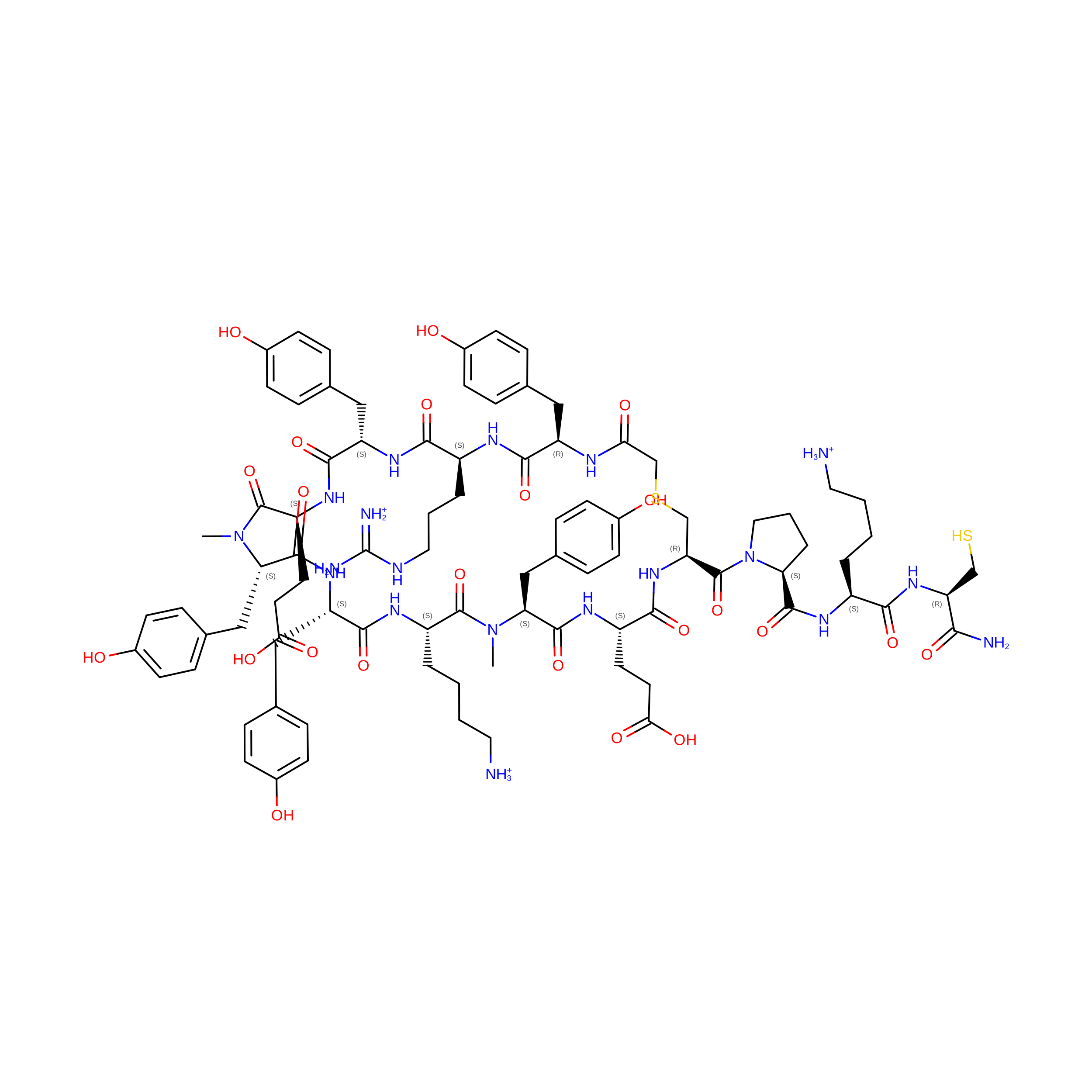
peptide Sa-D2E [auth a],
F [auth b],
G [auth c],
H [auth d]
15synthetic constructMutation(s): 0 
Entity Groups
Sequence Clusters30% Identity50% Identity70% Identity90% Identity95% Identity100% Identity
Sequence Annotations
Expand
Reference Sequence

Small Molecules

Ligands 3 Unique
IDChains Name / Formula / InChI Key2D Diagram3D Interactions
IMD

Query on IMD


Download:Ideal Coordinates CCD File
O [auth C],
S [auth D]
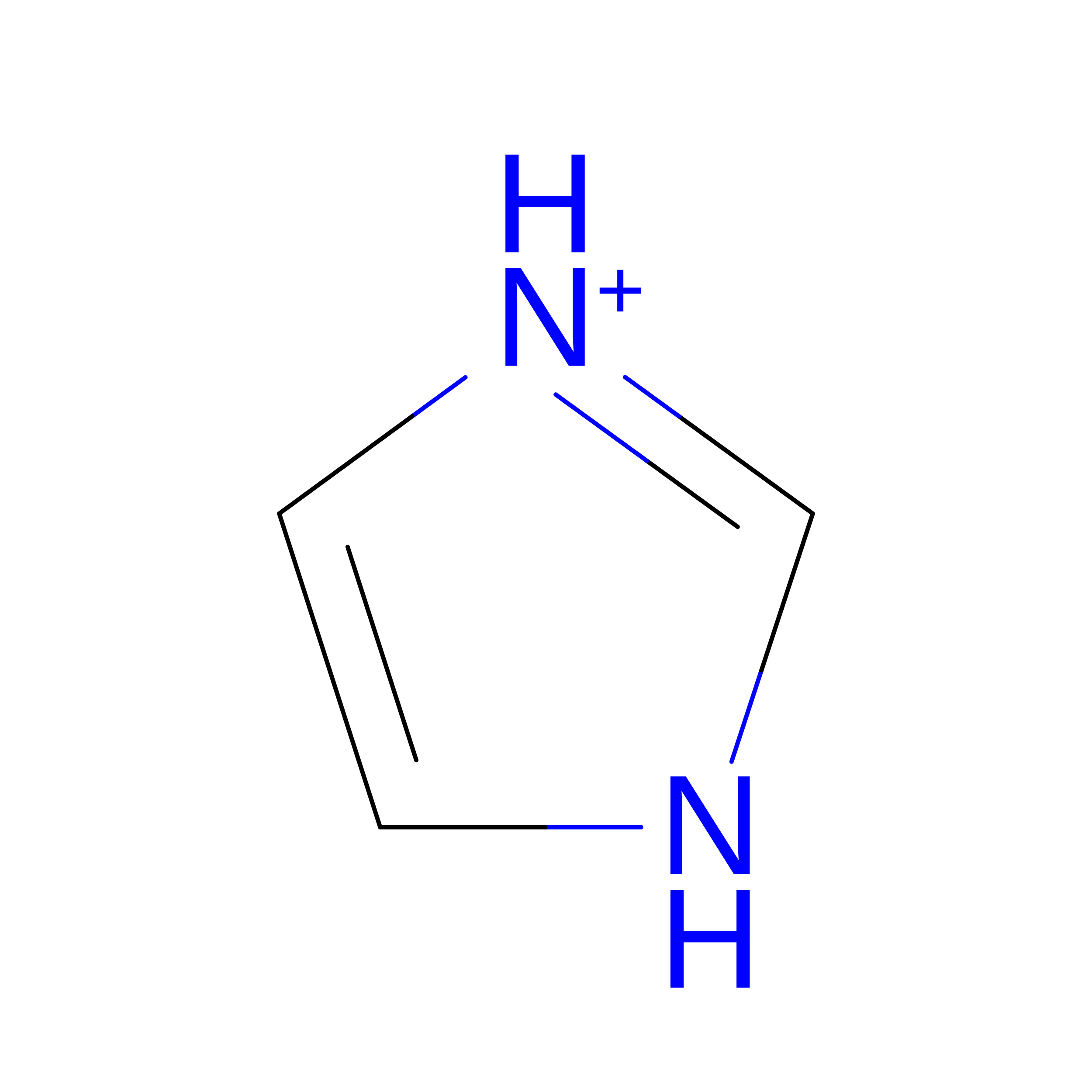
IMIDAZOLE
C3 H5 N2
RAXXELZNTBOGNW-UHFFFAOYSA-O
ZN
(Subject of Investigation/LOI)

Query on ZN


Download:Ideal Coordinates CCD File
I [auth A]
J [auth A]
L [auth B]
M [auth B]
P [auth C]
I [auth A],
J [auth A],
L [auth B],
M [auth B],
P [auth C],
Q [auth C],
T [auth D],
U [auth D]
ZINC ION
Zn
PTFCDOFLOPIGGS-UHFFFAOYSA-N
NA

Query on NA


Download:Ideal Coordinates CCD File
K [auth A],
N [auth B],
R [auth C],
V [auth D]
SODIUM ION
Na
FKNQFGJONOIPTF-UHFFFAOYSA-N
Modified Residues  2 Unique
IDChains TypeFormula2D DiagramParent
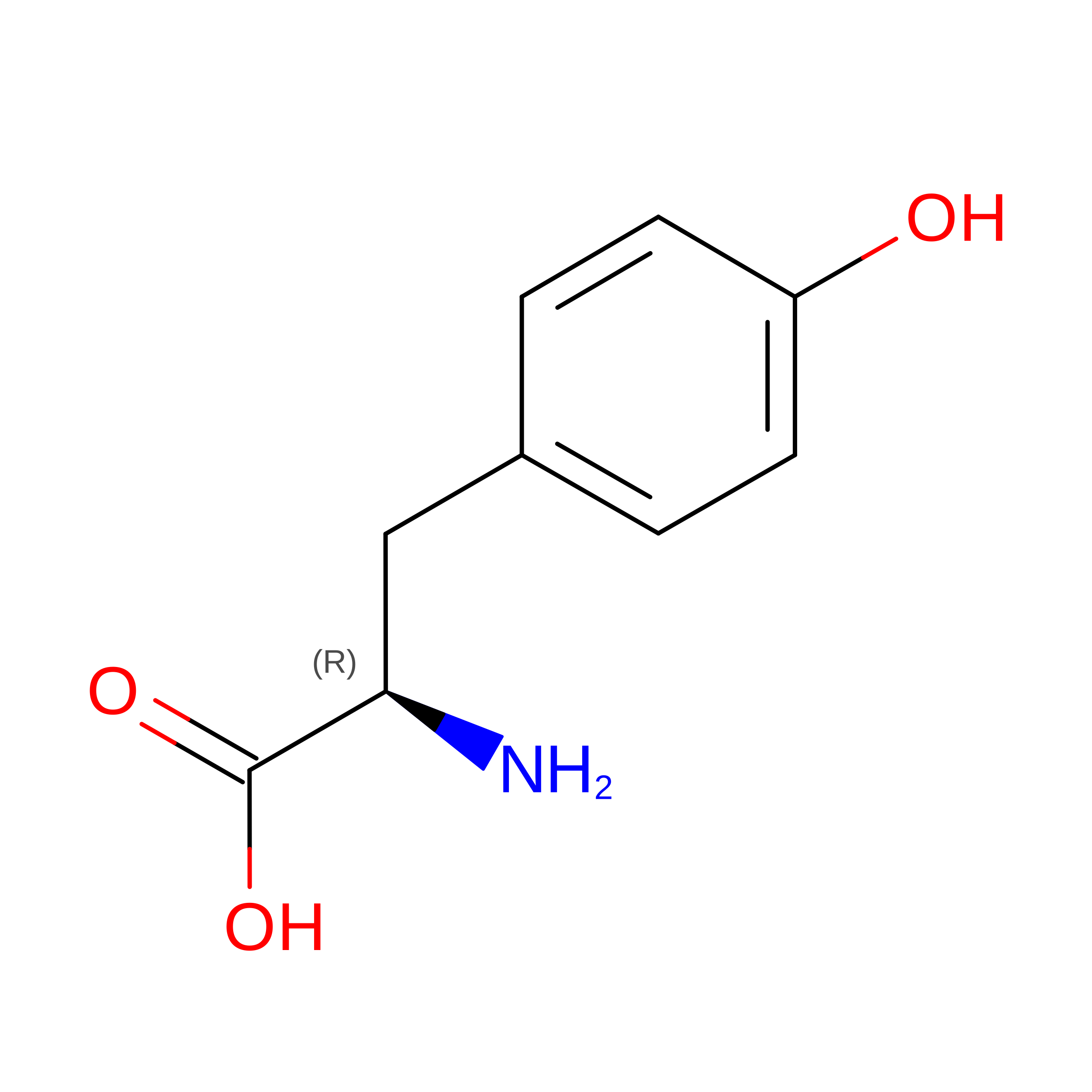
DTY
Query on DTY
E [auth a],
F [auth b],
G [auth c],
H [auth d]
D-PEPTIDE LINKINGC9 H11 N O3

--

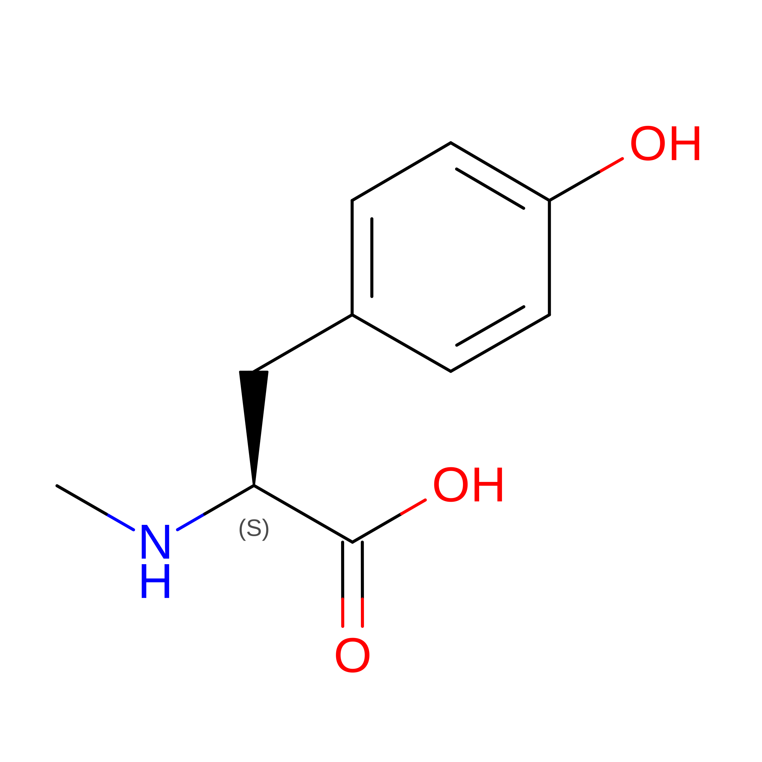
YNM
Query on YNM
E [auth a],
F [auth b],
G [auth c],
H [auth d]
L-PEPTIDE LINKINGC10 H13 N O3TYR

Experimental Data & Validation

Experimental Data

  • Method: X-RAY DIFFRACTION
  • Resolution: 1.90 Å
  • R-Value Free:  0.216 (Depositor), 0.221 (DCC) 
  • R-Value Work:  0.171 (Depositor), 0.178 (DCC) 
  • R-Value Observed: 0.173 (Depositor) 
Space Group: P 1 21 1
Unit Cell:
Length ( Å )Angle ( ˚ )
a = 77.999α = 90
b = 87.624β = 97.13
c = 151.341γ = 90
Software Package:
Software NamePurpose
XDSdata reduction
Aimlessdata scaling
PHASERphasing
PHENIXrefinement
PDB_EXTRACTdata extraction

Structure Validation

View Full Validation Report


Entry History 

& Funding Information

Deposition Data


Funding OrganizationLocationGrant Number
Not funded--

Revision History  (Full details and data files)

  • Version 1.0: 2022-08-10
    Type: Initial release
  • Version 1.1: 2022-08-31
    Changes: Database references
  • Version 1.2: 2023-10-18
    Changes: Data collection, Refinement description