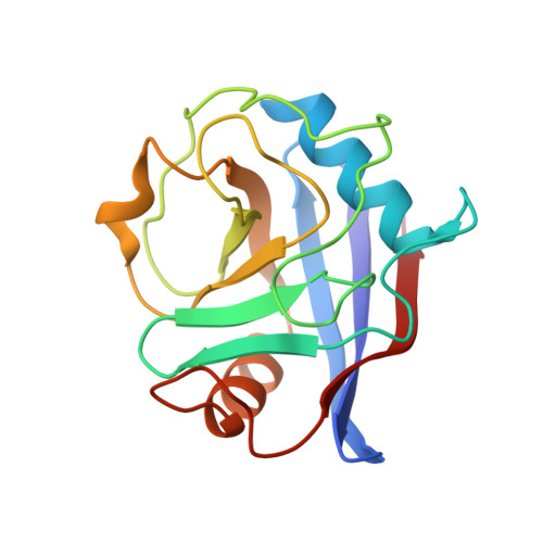
7THD

Structure of Cyclophilin D Peptidyl-Prolyl Isomerase Domain bound to Macrocyclic Inhibitor B52

  • Classification: ISOMERASE
  • Organism(s): Homo sapiens
  • Expression System: Escherichia coli BL21(DE3)
  • Mutation(s): Yes 

  • Deposited: 2022-01-10 Released: 2022-08-24 
  • Deposition Author(s): Rangwala, A.M., Thakur, M.K., Seeliger, M.A., Peterson, A.A., Liu, D.R.
  • Funding Organization(s): National Institutes of Health/National Cancer Institute (NIH/NCI), National Institutes of Health/National Institute of General Medical Sciences (NIH/NIGMS), National Institutes of Health/National Institute of Biomedical Imaging and Bioengineering (NIH/NIBIB), National Institutes of Health/Office of the Director, Howard Hughes Medical Institute (HHMI)

Experimental Data Snapshot

  • Method: X-RAY DIFFRACTION
  • Resolution: 1.16 Å
  • R-Value Free: 0.152 
  • R-Value Work: 0.136 
  • R-Value Observed: 0.136 

Starting Model: experimental
View more details

wwPDB Validation   3D Report Full Report


Ligand Structure Quality Assessment 


This is version 1.3 of the entry. See complete history


Literature

Discovery and molecular basis of subtype-selective cyclophilin inhibitors.

Peterson, A.A.Rangwala, A.M.Thakur, M.K.Ward, P.S.Hung, C.Outhwaite, I.R.Chan, A.I.Usanov, D.L.Mootha, V.K.Seeliger, M.A.Liu, D.R.

(2022) Nat Chem Biol 18: 1184-1195

  • DOI: https://doi.org/10.1038/s41589-022-01116-1
  • Primary Citation of Related Structures:  
    7TGS, 7TGT, 7TGU, 7TGV, 7TH1, 7TH6, 7TH7, 7THC, 7THD, 7THF

  • PubMed Abstract: 

    Although cyclophilins are attractive targets for probing biology and therapeutic intervention, no subtype-selective cyclophilin inhibitors have been described. We discovered novel cyclophilin inhibitors from the in vitro selection of a DNA-templated library of 256,000 drug-like macrocycles for cyclophilin D (CypD) affinity. Iterated macrocycle engineering guided by ten X-ray co-crystal structures yielded potent and selective inhibitors (half maximal inhibitory concentration (IC 50 ) = 10 nM) that bind the active site of CypD and also make novel interactions with non-conserved residues in the S2 pocket, an adjacent exo-site. The resulting macrocycles inhibit CypD activity with 21- to >10,000-fold selectivity over other cyclophilins and inhibit mitochondrial permeability transition pore opening in isolated mitochondria. We further exploited S2 pocket interactions to develop the first cyclophilin E (CypE)-selective inhibitor, which forms a reversible covalent bond with a CypE S2 pocket lysine, and exhibits 30- to >4,000-fold selectivity over other cyclophilins. These findings reveal a strategy to generate isoform-selective small-molecule cyclophilin modulators, advancing their suitability as targets for biological investigation and therapeutic development.


  • Organizational Affiliation

    Merkin Institute of Transformative Technologies in Healthcare, Broad Institute of MIT and Harvard, Cambridge, MA, USA.


Macromolecules
Find similar proteins by:  (by identity cutoff)  |  3D Structure
Entity ID: 1
MoleculeChains Sequence LengthOrganismDetailsImage
Peptidyl-prolyl cis-trans isomerase F, mitochondrial163Homo sapiensMutation(s): 1 
Gene Names: PPIFCYP3
EC: 5.2.1.8
UniProt & NIH Common Fund Data Resources
Find proteins for P30405 (Homo sapiens)
Explore P30405 
Go to UniProtKB:  P30405
PHAROS:  P30405
GTEx:  ENSG00000108179 
Entity Groups  
Sequence Clusters30% Identity50% Identity70% Identity90% Identity95% Identity100% Identity
UniProt GroupP30405
Sequence Annotations
Expand
  • Reference Sequence
Small Molecules
Ligands 1 Unique
IDChains Name / Formula / InChI Key2D Diagram3D Interactions
I4V (Subject of Investigation/LOI)
Query on I4V

Download Ideal Coordinates CCD File 
B [auth A][(4'-{[(4S,7S,11R,13E,19S)-19-{[2-(2-aminoethoxy)ethyl]carbamoyl}-7-benzyl-3,6,12,15,21-pentaoxo-1,3,4,5,6,7,8,9,10,12,15,16,17,18,19,20,21,22-octadecahydro-2H-7,11-methano-2,5,11,16,20-benzopentaazacyclotetracosin-4-yl]methyl}[1,1'-biphenyl]-4-yl)methyl]propanedioic acid
C53 H61 N7 O11
ZHNYUSCORILKFS-FCZRSVTRSA-N
Experimental Data & Validation

Experimental Data

  • Method: X-RAY DIFFRACTION
  • Resolution: 1.16 Å
  • R-Value Free: 0.152 
  • R-Value Work: 0.136 
  • R-Value Observed: 0.136 
  • Space Group: P 2 21 21
Unit Cell:
Length ( Å )Angle ( ˚ )
a = 38.408α = 90
b = 67.032β = 90
c = 69.324γ = 90
Software Package:
Software NamePurpose
PHENIXrefinement
Cootmodel building
XDSdata reduction
autoPROCdata scaling
autoPROCdata processing
PHASERphasing

Structure Validation

View Full Validation Report


Ligand Structure Quality Assessment 


Entry History & Funding Information

Deposition Data


Funding OrganizationLocationGrant Number
National Institutes of Health/National Cancer Institute (NIH/NCI)United StatesCA260772
National Institutes of Health/National Institute of General Medical Sciences (NIH/NIGMS)United StatesGM118062
National Institutes of Health/National Institute of Biomedical Imaging and Bioengineering (NIH/NIBIB)United StatesEB022376
National Institutes of Health/National Institute of General Medical Sciences (NIH/NIGMS)United StatesGM119437
National Institutes of Health/Office of the DirectorUnited StatesOD028478
Howard Hughes Medical Institute (HHMI)United States--

Revision History  (Full details and data files)

  • Version 1.0: 2022-08-24
    Type: Initial release
  • Version 1.1: 2022-10-12
    Changes: Database references
  • Version 1.2: 2022-11-02
    Changes: Database references
  • Version 1.3: 2023-10-18
    Changes: Data collection, Refinement description