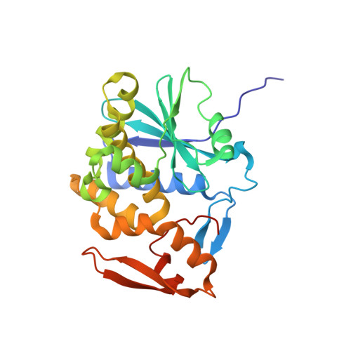
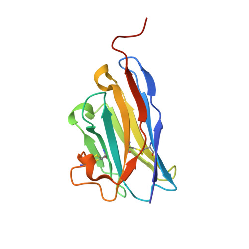
7TH2

Single-domain VHH intrabodies neutralize ricin toxin


Experimental Data Snapshot

  • Method: X-RAY DIFFRACTION
  • Resolution: 1.84 Å
  • R-Value Free: 0.187 
  • R-Value Work: 0.149 
  • R-Value Observed: 0.151 

Starting Models: experimental
View more details

wwPDB Validation   3D Report Full Report


This is version 1.2 of the entry. See complete history


Literature

Single-domain antibodies neutralize ricin toxin intracellularly by blocking access to ribosomal P-stalk proteins.

Czajka, T.F.Vance, D.J.Davis, S.Rudolph, M.J.Mantis, N.J.

(2022) J Biol Chem 298: 101742-101742

  • DOI: https://doi.org/10.1016/j.jbc.2022.101742
  • Primary Citation of Related Structures:  
    7TGF, 7TGI, 7TH2, 7TH3

  • PubMed Abstract: 

    During ricin intoxication in mammalian cells, ricin's enzymatic (RTA) and binding (RTB) subunits disassociate in the endoplasmic reticulum. RTA is then translocated into the cytoplasm where, by virtue of its ability to depurinate a conserved residue within the sarcin-ricin loop (SRL) of 28S rRNA, it functions as a ribosome-inactivating protein. It has been proposed that recruitment of RTA to the SRL is facilitated by ribosomal P-stalk proteins, whose C-terminal domains interact with a cavity on RTA normally masked by RTB; however, evidence that this interaction is critical for RTA activity within cells is lacking. Here, we characterized a collection of single-domain antibodies (V H Hs) whose epitopes overlap with the P-stalk binding pocket on RTA. The crystal structures of three such V H Hs (V9E1, V9F9, and V9B2) in complex with RTA revealed not only occlusion of the ribosomal P-stalk binding pocket but also structural mimicry of C-terminal domain peptides by complementarity-determining region 3. In vitro assays confirmed that these V H Hs block RTA-P-stalk peptide interactions and protect ribosomes from depurination. Moreover, when expressed as "intrabodies," these V H Hs rendered cells resistant to ricin intoxication. One V H H (V9F6), whose epitope was structurally determined to be immediately adjacent to the P-stalk binding pocket, was unable to neutralize ricin within cells or protect ribosomes from RTA in vitro. These findings are consistent with the recruitment of RTA to the SRL by ribosomal P-stalk proteins as a requisite event in ricin-induced ribosome inactivation.


  • Organizational Affiliation

    Department of Biomedical Sciences, University at Albany, Albany, New York, USA.


Macromolecules
Find similar proteins by:  (by identity cutoff)  |  3D Structure
Entity ID: 1
MoleculeChains Sequence LengthOrganismDetailsImage
Ricin A chain
A, B
269Ricinus communisMutation(s): 0 
EC: 3.2.2.22
UniProt
Find proteins for P02879 (Ricinus communis)
Explore P02879 
Go to UniProtKB:  P02879
Entity Groups  
Sequence Clusters30% Identity50% Identity70% Identity90% Identity95% Identity100% Identity
UniProt GroupP02879
Sequence Annotations
Expand
  • Reference Sequence
Find similar proteins by:  (by identity cutoff)  |  3D Structure
Entity ID: 2
MoleculeChains Sequence LengthOrganismDetailsImage
VHH antibody
C, D
132Vicugna pacosMutation(s): 0 
Entity Groups  
Sequence Clusters30% Identity50% Identity70% Identity90% Identity95% Identity100% Identity
Sequence Annotations
Expand
  • Reference Sequence
Small Molecules
Ligands 4 Unique
IDChains Name / Formula / InChI Key2D Diagram3D Interactions
MES
Query on MES

Download Ideal Coordinates CCD File 
N [auth A],
Z [auth B]
2-(N-MORPHOLINO)-ETHANESULFONIC ACID
C6 H13 N O4 S
SXGZJKUKBWWHRA-UHFFFAOYSA-N
SO4
Query on SO4

Download Ideal Coordinates CCD File 
CA [auth C],
X [auth B],
Y [auth B]
SULFATE ION
O4 S
QAOWNCQODCNURD-UHFFFAOYSA-L
EDO
Query on EDO

Download Ideal Coordinates CCD File 
EA [auth D],
FA [auth D]
1,2-ETHANEDIOL
C2 H6 O2
LYCAIKOWRPUZTN-UHFFFAOYSA-N
CL
Query on CL

Download Ideal Coordinates CCD File 
AA [auth C]
BA [auth C]
DA [auth D]
E [auth A]
F [auth A]
AA [auth C],
BA [auth C],
DA [auth D],
E [auth A],
F [auth A],
G [auth A],
H [auth A],
I [auth A],
J [auth A],
K [auth A],
L [auth A],
M [auth A],
O [auth B],
P [auth B],
Q [auth B],
R [auth B],
S [auth B],
T [auth B],
U [auth B],
V [auth B],
W [auth B]
CHLORIDE ION
Cl
VEXZGXHMUGYJMC-UHFFFAOYSA-M
Experimental Data & Validation

Experimental Data

  • Method: X-RAY DIFFRACTION
  • Resolution: 1.84 Å
  • R-Value Free: 0.187 
  • R-Value Work: 0.149 
  • R-Value Observed: 0.151 
  • Space Group: P 1 21 1
Unit Cell:
Length ( Å )Angle ( ˚ )
a = 60.644α = 90
b = 63.812β = 107.12
c = 99.298γ = 90
Software Package:
Software NamePurpose
SCALEPACKdata scaling
PHENIXrefinement
PDB_EXTRACTdata extraction
HKL-2000data reduction
PHASERphasing

Structure Validation

View Full Validation Report


Entry History & Funding Information

Deposition Data


Funding OrganizationLocationGrant Number
National Institutes of Health/National Institute Of Allergy and Infectious Diseases (NIH/NIAID)United StatesHHSN272201400021C

Revision History  (Full details and data files)

  • Version 1.0: 2022-10-12
    Type: Initial release
  • Version 1.1: 2023-10-18
    Changes: Data collection, Refinement description
  • Version 1.2: 2024-10-23
    Changes: Structure summary