7TEY | pdb_00007tey

Cryo-EM structure of SARS-CoV-2 Delta (B.1.617.2) spike protein


Experimental Data Snapshot

  • Method: ELECTRON MICROSCOPY
  • Resolution: 2.25 Å
  • Aggregation State: PARTICLE 
  • Reconstruction Method: SINGLE PARTICLE 

wwPDB Validation 3D Report Full Report

Validation slider image for 7TEY

This is version 1.1 of the entry. See complete history

Literature

Structural and biochemical rationale for enhanced spike protein fitness in delta and kappa SARS-CoV-2 variants.

Saville, J.W.Mannar, D.Zhu, X.Srivastava, S.S.Berezuk, A.M.Demers, J.P.Zhou, S.Tuttle, K.S.Sekirov, I.Kim, A.Li, W.Dimitrov, D.S.Subramaniam, S.

(2022) Nat Commun 13: 742-742

  • DOI: https://doi.org/10.1038/s41467-022-28324-6
  • Primary Citation Related Structures: 
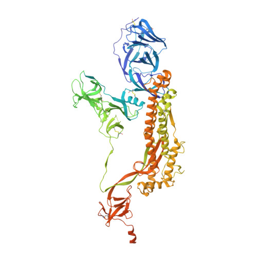
    7TEW, 7TEX, 7TEY, 7TEZ, 7TF0, 7TF1, 7TF2, 7TF3, 7TF4, 7TF5

  • PubMed Abstract: 

    The Delta and Kappa variants of SARS-CoV-2 co-emerged in India in late 2020, with the Delta variant underlying the resurgence of COVID-19, even in countries with high vaccination rates. In this study, we assess structural and biochemical aspects of viral fitness for these two variants using cryo-electron microscopy (cryo-EM), ACE2-binding and antibody neutralization analyses. Both variants demonstrate escape of antibodies targeting the N-terminal domain, an important immune hotspot for neutralizing epitopes. Compared to wild-type and Kappa lineages, Delta variant spike proteins show modest increase in ACE2 affinity, likely due to enhanced electrostatic complementarity at the RBD-ACE2 interface, which we characterize by cryo-EM. Unexpectedly, Kappa variant spike trimers form a structural head-to-head dimer-of-trimers assembly, which we demonstrate is a result of the E484Q mutation and with unknown biological implications. The combination of increased antibody escape and enhanced ACE2 binding provides an explanation, in part, for the rapid global dominance of the Delta variant.


  • Organizational Affiliation
    • Department of Biochemistry and Molecular Biology, University of British Columbia, Vancouver, BC, V6T 1Z3, Canada.

Macromolecule Content 

  • Total Structure Weight: 438.6 kDa 
  • Atom Count: 21,867 
  • Modeled Residue Count: 2,703 
  • Deposited Residue Count: 3,858 
  • Unique protein chains: 1

Macromolecules

Find similar proteins by:|  3D Structure
Entity ID: 1
MoleculeChains  Sequence LengthOrganismDetailsImage
Spike glycoprotein
A, B, C
1,286Severe acute respiratory syndrome coronavirus 2Mutation(s): 16 
Gene Names: S2
UniProt
Find proteins for P0DTC2 (Severe acute respiratory syndrome coronavirus 2)
Explore P0DTC2 
Go to UniProtKB:  P0DTC2
Entity Groups
Sequence Clusters30% Identity50% Identity70% Identity90% Identity95% Identity100% Identity
UniProt GroupP0DTC2
Glycosylation
Glycosylation Sites: 12Go to GlyGen: P0DTC2-1
Sequence Annotations
Expand
Reference Sequence

Small Molecules

Ligands 1 Unique
IDChains Name / Formula / InChI Key2D Diagram3D Interactions
NAG

Query on NAG


Download:Ideal Coordinates CCD File
AA [auth A]
BA [auth B]
CA [auth B]
DA [auth B]
EA [auth B]
AA [auth A],
BA [auth B],
CA [auth B],
DA [auth B],
EA [auth B],
FA [auth B],
GA [auth B],
HA [auth C],
IA [auth C],
JA [auth C],
KA [auth C],
LA [auth C],
MA [auth C],
NA [auth C],
OA [auth C],
V [auth A],
W [auth A],
X [auth A],
Y [auth A],
Z [auth A]
2-acetamido-2-deoxy-beta-D-glucopyranose
C8 H15 N O6
OVRNDRQMDRJTHS-FMDGEEDCSA-N

Experimental Data & Validation

Experimental Data

  • Method: ELECTRON MICROSCOPY
  • Resolution: 2.25 Å
  • Aggregation State: PARTICLE 
  • Reconstruction Method: SINGLE PARTICLE 

Structure Validation

View Full Validation Report


Entry History 

& Funding Information

Deposition Data


Funding OrganizationLocationGrant Number
Canada Excellence Research Chair AwardPrecision Cancer Drug Design
Other governmentCOVID-19 research

Revision History  (Full details and data files)

  • Version 1.0: 2022-03-16
    Type: Initial release
  • Version 1.1: 2024-10-09
    Changes: Data collection, Structure summary