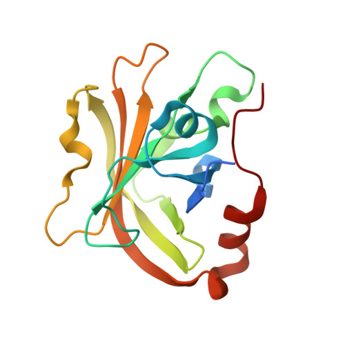
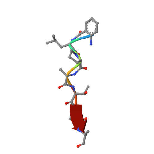
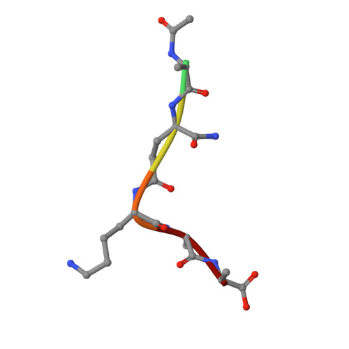
7T8Z

Structure of Class A sortase from Streptococcus pyogenes bound to lipid II mimetic, LPATA: Thr-out conformation


Experimental Data Snapshot

  • Method: X-RAY DIFFRACTION
  • Resolution: 1.90 Å
  • R-Value Free: 0.229 
  • R-Value Work: 0.184 
  • R-Value Observed: 0.186 

Starting Model: experimental
View more details

wwPDB Validation   3D Report Full Report


This is version 2.0 of the entry. See complete history


Literature

Structures of Streptococcus pyogenes class A sortase in complex with substrate and product mimics provide key details of target recognition.

Johnson, D.A.Piper, I.M.Vogel, B.A.Jackson, S.N.Svendsen, J.E.Kodama, H.M.Lee, D.E.Lindblom, K.M.McCarty, J.Antos, J.M.Amacher, J.F.

(2022) J Biol Chem 298: 102446-102446

  • DOI: https://doi.org/10.1016/j.jbc.2022.102446
  • Primary Citation of Related Structures:  
    7S4O, 7S51, 7T8Y, 7T8Z

  • PubMed Abstract: 

    The cell wall is a critical extracellular barrier for bacteria and many other organisms. In bacteria, this structural layer consists of peptidoglycan, which maintains cell shape and structural integrity and provides a scaffold for displaying various protein factors. To attach proteins to the cell wall, Gram-positive bacteria utilize sortase enzymes, which are cysteine transpeptidases that recognize and cleave a specific sorting signal, followed by ligation of the sorting signal-containing protein to the peptidoglycan precursor lipid II (LII). This mechanism is the subject of considerable interest as a target for therapeutic intervention and as a tool for protein engineering, where sortases have enabled sortase-mediated ligation or sortagging strategies. Despite these uses, there remains an incomplete understanding of the stereochemistry of substrate recognition and ligation product formation. Here, we solved the first structures of sortase A from Streptococcus pyogenes bound to two substrate sequences, LPATA and LPATS. In addition, we synthesized a mimetic of the product of sortase-mediated ligation involving LII (LPAT-LII) and solved the complex structure in two ligand conformations. These structures were further used as the basis for molecular dynamics simulations to probe sortase A-ligand dynamics and to construct a model of the acyl-enzyme intermediate, thus providing a structural view of multiple key states in the catalytic mechanism. Overall, this structural information provides new insights into the recognition of the sortase substrate motif and LII ligation partner and will support the continued development of sortases for protein engineering applications.


  • Organizational Affiliation

    Department of Chemistry, Western Washington University, Bellingham, Washington, USA.


Macromolecules
Find similar proteins by:  (by identity cutoff)  |  3D Structure
Entity ID: 1
MoleculeChains Sequence LengthOrganismDetailsImage
Sortase170Streptococcus pyogenesMutation(s): 0 
Gene Names: 
UniProt
Find proteins for Q99ZN4 (Streptococcus pyogenes serotype M1)
Explore Q99ZN4 
Go to UniProtKB:  Q99ZN4
Entity Groups  
Sequence Clusters30% Identity50% Identity70% Identity90% Identity95% Identity100% Identity
UniProt GroupQ99ZN4
Sequence Annotations
Expand
  • Reference Sequence

Find similar proteins by:  Sequence   |   3D Structure  

Entity ID: 2
MoleculeChains Sequence LengthOrganismDetailsImage
BE2-LEU-PRO-ALA-THR-ALA-ALA7synthetic constructMutation(s): 0 
Sequence Annotations
Expand
  • Reference Sequence

Find similar proteins by:  Sequence   |   3D Structure  

Entity ID: 3
MoleculeChains Sequence LengthOrganismDetailsImage
ACE-ALA-ZGL-LYS-DAL-DALC [auth D]6synthetic constructMutation(s): 0 
Sequence Annotations
Expand
  • Reference Sequence
Biologically Interesting Molecules (External Reference) 2 Unique
Experimental Data & Validation

Experimental Data

  • Method: X-RAY DIFFRACTION
  • Resolution: 1.90 Å
  • R-Value Free: 0.229 
  • R-Value Work: 0.184 
  • R-Value Observed: 0.186 
  • Space Group: P 21 21 21
Unit Cell:
Length ( Å )Angle ( ˚ )
a = 34.324α = 90
b = 57.677β = 90
c = 71.457γ = 90
Software Package:
Software NamePurpose
XDSdata reduction
Aimlessdata scaling
PHENIXrefinement
PDB_EXTRACTdata extraction
PHENIXphasing

Structure Validation

View Full Validation Report


Entry History & Funding Information

Deposition Data


Funding OrganizationLocationGrant Number
National Science Foundation (NSF, United States)United States2044958

Revision History  (Full details and data files)

  • Version 1.0: 2022-09-07
    Type: Initial release
  • Version 1.1: 2022-09-14
    Changes: Database references
  • Version 1.2: 2022-10-12
    Changes: Database references
  • Version 1.3: 2023-10-18
    Changes: Data collection, Refinement description
  • Version 2.0: 2023-11-15
    Changes: Atomic model, Data collection, Derived calculations