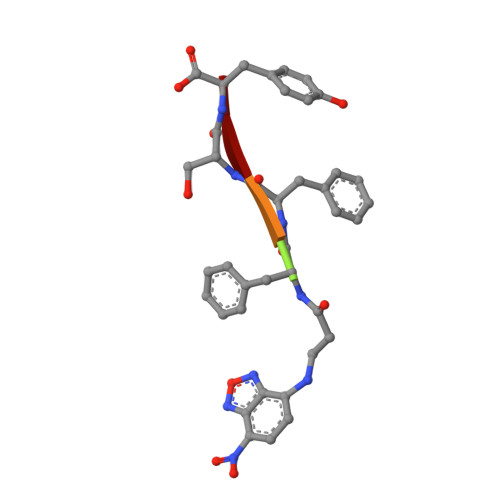
Cell spheroid creation by transcytotic intercellular gelation.
Guo, J., Wang, F., Huang, Y., He, H., Tan, W., Yi, M., Egelman, E.H., Xu, B.(2023) Nat Nanotechnol 18: 1094-1104
- PubMed: 37217766
- DOI: https://doi.org/10.1038/s41565-023-01401-7
- Primary Citation of Related Structures:
7T6E, 8DST, 8FOF - PubMed Abstract:
Cell spheroids bridge the discontinuity between in vitro systems and in vivo animal models. However, inducing cell spheroids by nanomaterials remains an inefficient and poorly understood process. Here we use cryogenic electron microscopy to determine the atomic structure of helical nanofibres self-assembled from enzyme-responsive D-peptides and fluorescent imaging to show that the transcytosis of D-peptides induces intercellular nanofibres/gels that potentially interact with fibronectin to enable cell spheroid formation. Specifically, D-phosphopeptides, being protease resistant, undergo endocytosis and endosomal dephosphorylation to generate helical nanofibres. On secretion to the cell surface, these nanofibres form intercellular gels that act as artificial matrices and facilitate the fibrillogenesis of fibronectins to induce cell spheroids. No spheroid formation occurs without endo- or exocytosis, phosphate triggers or shape switching of the peptide assemblies. This study-coupling transcytosis and morphological transformation of peptide assemblies-demonstrates a potential approach for regenerative medicine and tissue engineering.
- Department of Chemistry, Brandeis University, Waltham, MA, USA.
Organizational Affiliation:


