7T1O | pdb_00007t1o

Solution structure of 7SK stem-loop 1 with HIV-1 Tat Subtype G Arginine Rich Motif

  • Classification: TRANSCRIPTION
  • Organism(s): Homo sapiens, HIV-1 06TG.HT008
  • Mutation(s): No 

  • Deposited: 2021-12-02 Released: 2022-09-14 
  • Deposition Author(s): Pham, V.V., Gao, M., D'Souza, V.M.
  • Funding Organization(s): Howard Hughes Medical Institute (HHMI), National Institutes of Health/National Institute Of Allergy and Infectious Diseases (NIH/NIAID)

Experimental Data Snapshot

  • Method: SOLUTION NMR
  • Conformers Calculated: 100 
  • Conformers Submitted: 10 
  • Selection Criteria: structures with the lowest energy 

  • Method: SOLUTION SCATTERING

wwPDB Validation 3D Report Full Report

Validation slider image for 7T1O

This is version 1.1 of the entry. See complete history

Literature

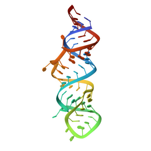
A structure-based mechanism for displacement of the HEXIM adapter from 7SK small nuclear RNA.

Pham, V.V.Gao, M.Meagher, J.L.Smith, J.L.D'Souza, V.M.

(2022) Commun Biol 5: 819-819

  • DOI: https://doi.org/10.1038/s42003-022-03734-w
  • Primary Citation Related Structures: 
    7T1N, 7T1O, 7T1P

  • PubMed Abstract: 

    Productive transcriptional elongation of many cellular and viral mRNAs requires transcriptional factors to extract pTEFb from the 7SK snRNP by modulating the association between HEXIM and 7SK snRNA. In HIV-1, Tat binds to 7SK by displacing HEXIM. However, without the structure of the 7SK-HEXIM complex, the constraints that must be overcome for displacement remain unknown. Furthermore, while structure details of the Tat NL4-3 -7SK complex have been elucidated, it is unclear how subtypes with more HEXIM-like Tat sequences accomplish displacement. Here we report the structures of HEXIM, Tat G , and Tat Fin arginine rich motifs in complex with the apical stemloop-1 of 7SK. While most interactions between 7SK with HEXIM and Tat are similar, critical differences exist that guide function. First, the conformational plasticity of 7SK enables the formation of three different base pair configurations at a critical remodeling site, which allows for the modulation required for HEXIM binding and its subsequent displacement by Tat. Furthermore, the specific sequence variations observed in various Tat subtypes all converge on remodeling 7SK at this region. Second, we show that HEXIM primes its own displacement by causing specific local destabilization upon binding - a feature that is then exploited by Tat to bind 7SK more efficiently.


  • Organizational Affiliation
    • Department of Molecular and Cellular Biology, Harvard University, Cambridge, MA, 02138, USA.

Macromolecule Content 

  • Total Structure Weight: 20.16 kDa 
  • Atom Count: 1,340 
  • Modeled Residue Count: 73 
  • Deposited Residue Count: 73 
  • Unique protein chains: 1
  • Unique nucleic acid chains: 1

Macromolecules


Find similar proteins by:  Sequence   |   3D Structure  
Entity ID: 2
MoleculeChains  Sequence LengthOrganismDetailsImage
Tat Subtype G Arginine Rich Motif17HIV-1 06TG.HT008Mutation(s): 0 
Find similar nucleic acids by:  (by identity cutoff) 
Entity ID: 1
MoleculeChains LengthOrganismImage
7SK stem-loop 1 RNA (56-MER)56Homo sapiens
Sequence Annotations
Expand
Reference Sequence

Experimental Data & Validation

Experimental Data

  • Method: SOLUTION NMR
  • Conformers Calculated: 100 
  • Conformers Submitted: 10 
  • Selection Criteria: structures with the lowest energy 
  • Method: SOLUTION SCATTERING

Structure Validation

View Full Validation Report


Entry History 

& Funding Information

Deposition Data


Funding OrganizationLocationGrant Number
Howard Hughes Medical Institute (HHMI)United States55108516
National Institutes of Health/National Institute Of Allergy and Infectious Diseases (NIH/NIAID)United StatesU54 AI50470

Revision History  (Full details and data files)

  • Version 1.0: 2022-09-14
    Type: Initial release
  • Version 1.1: 2024-05-15
    Changes: Data collection, Database references