


Development of PRPK Directed Phthalimides
Seo, H.-S., Mizutani, T., Hideshima, T., Vangos, N.E., Zhang, T., Anderson, K.C., Gray, N.S., Dhe-Paganon, S.(2021) bioRxiv
Experimental Data Snapshot
Starting Models: experimental
View more details
(2021) bioRxiv
Entity ID: 1 | |||||
|---|---|---|---|---|---|
| Molecule | Chains | Sequence Length | Organism | Details | Image |
| EKC/KEOPS complex subunit TP53RK | 255 | Homo sapiens | Mutation(s): 0 Gene Names: TP53RK, C20orf64, PRPK EC: 3.6 (PDB Primary Data), 2.7.11.1 (PDB Primary Data) | 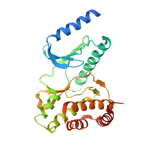 | |
UniProt & NIH Common Fund Data Resources | |||||
PHAROS: Q96S44 GTEx: ENSG00000172315 | |||||
Entity Groups | |||||
| Sequence Clusters | 30% Identity50% Identity70% Identity90% Identity95% Identity100% Identity | ||||
| UniProt Group | Q96S44 | ||||
Sequence AnnotationsExpand | |||||
Reference Sequence | |||||
Entity ID: 2 | |||||
|---|---|---|---|---|---|
| Molecule | Chains | Sequence Length | Organism | Details | Image |
| EKC/KEOPS complex subunit TPRKB | 178 | Homo sapiens | Mutation(s): 0 Gene Names: TPRKB, CGI-121, My019 | 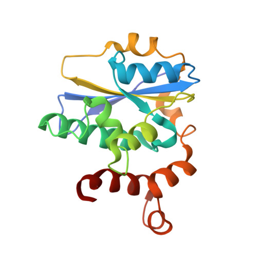 | |
UniProt & NIH Common Fund Data Resources | |||||
PHAROS: Q9Y3C4 GTEx: ENSG00000144034 | |||||
Entity Groups | |||||
| Sequence Clusters | 30% Identity50% Identity70% Identity90% Identity95% Identity100% Identity | ||||
| UniProt Group | Q9Y3C4 | ||||
Sequence AnnotationsExpand | |||||
Reference Sequence | |||||
| Ligands 1 Unique | |||||
|---|---|---|---|---|---|
| ID | Chains | Name / Formula / InChI Key | 2D Diagram | 3D Interactions | |
| DVO (Subject of Investigation/LOI) Download:Ideal Coordinates CCD File | E [auth A], F [auth C] | 2-[(3R)-3-methyl-2,6-dioxopiperidin-3-yl]-1,3-dioxo-2,3-dihydro-1H-isoindole-5-carboxylic acid C15 H12 N2 O6 VWVJRXADTDRXRW-OAHLLOKOSA-N |  | ||
| Length ( Å ) | Angle ( ˚ ) |
|---|---|
| a = 65.578 | α = 90 |
| b = 79.038 | β = 105.614 |
| c = 100.889 | γ = 90 |
| Software Name | Purpose |
|---|---|
| PHENIX | refinement |
| PHENIX | refinement |
| XDS | data reduction |
| Aimless | data scaling |
| PHASER | phasing |
| PDB_EXTRACT | data extraction |
| Funding Organization | Location | Grant Number |
|---|---|---|
| National Institutes of Health/National Cancer Institute (NIH/NCI) | United States | -- |


