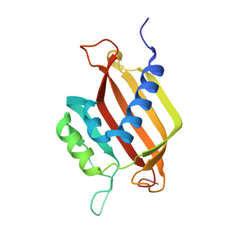
7SUO

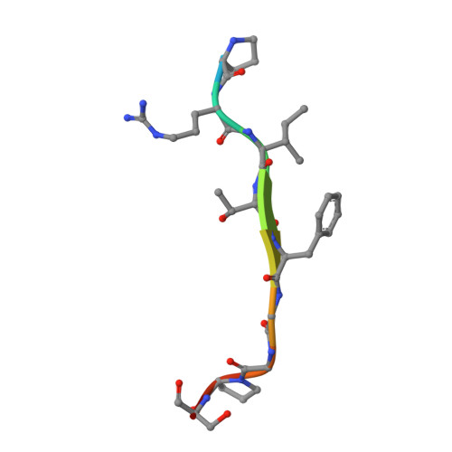
Crystal Structure of the G3BP1 NTF2-like domain bound to the IDR1 of SARS-CoV-2 nucleocapsid protein

  • Classification: Hydrolase/Viral Protein
  • Organism(s): Homo sapiens, Severe acute respiratory syndrome coronavirus 2
  • Expression System: Escherichia coli
  • Mutation(s): No 

  • Deposited: 2021-11-17 Released: 2022-03-16 
  • Deposition Author(s): Biswal, M., Lu, J., Song, J.
  • Funding Organization(s): National Institutes of Health/National Library of Medicine (NIH/NLM), National Institutes of Health/National Institute of General Medical Sciences (NIH/NIGMS), National Institutes of Health/National Institute Of Allergy and Infectious Diseases (NIH/NIAID), National Institutes of Health/Eunice Kennedy Shriver National Institute of Child Health & Human Development (NIH/NICHD), National Institutes of Health/National Institute of Environmental Health Sciences (NIH/NIEHS)

Experimental Data Snapshot

  • Method: X-RAY DIFFRACTION
  • Resolution: 2.35 Å
  • R-Value Free: 0.248 
  • R-Value Work: 0.197 
  • R-Value Observed: 0.202 

Starting Model: experimental
View more details

wwPDB Validation   3D Report Full Report


This is version 1.2 of the entry. See complete history


Literature

SARS-CoV-2 Nucleocapsid Protein Targets a Conserved Surface Groove of the NTF2-like Domain of G3BP1.

Biswal, M.Lu, J.Song, J.

(2022) J Mol Biol 434: 167516-167516

  • DOI: https://doi.org/10.1016/j.jmb.2022.167516
  • Primary Citation of Related Structures:  
    7SUO

  • PubMed Abstract: 

    Stress granule (SG) formation mediated by Ras GTPase-activating protein-binding protein 1 (G3BP1) constitutes a key obstacle for viral replication, which makes G3BP1 a frequent target for viruses. For instance, the SARS-CoV-2 nucleocapsid (N) protein interacts with G3BP1 directly to suppress SG assembly and promote viral production. However, the molecular basis for the SARS-CoV-2 N - G3BP1 interaction remains elusive. Here we report biochemical and structural analyses of the SARS-CoV-2 N - G3BP1 interaction, revealing differential contributions of various regions of SARS-CoV-2 N to G3BP1 binding. The crystal structure of the NTF2-like domain of G3BP1 (G3BP1 NTF2 ) in complex with a peptide derived from SARS-CoV-2 N (residues 1-25, N 1-25 ) reveals that SARS-CoV-2 N 1-25 occupies a conserved surface groove of G3BP1 NTF2 via surface complementarity. We show that a φ-x-F (φ, hydrophobic residue) motif constitutes the primary determinant for G3BP1 NTF2 -targeting proteins, while the flanking sequence underpins diverse secondary interactions. We demonstrate that mutation of key interaction residues of the SARS-CoV-2 N 1-25  - G3BP1 NTF2 complex leads to disruption of the SARS-CoV-2 N - G3BP1 interaction in vitro. Together, these results provide a molecular basis of the strain-specific interaction between SARS-CoV-2 N and G3BP1, which has important implications for the development of novel therapeutic strategies against SARS-CoV-2 infection.


  • Organizational Affiliation

    Department of Biochemistry, University of California, Riverside, CA, USA.


Macromolecules
Find similar proteins by:  (by identity cutoff)  |  3D Structure
Entity ID: 1
MoleculeChains Sequence LengthOrganismDetailsImage
Ras GTPase-activating protein-binding protein 1
A, B
138Homo sapiensMutation(s): 0 
Gene Names: G3BP1G3BP
EC: 3.6.4.12 (PDB Primary Data), 3.6.4.13 (PDB Primary Data)
UniProt & NIH Common Fund Data Resources
Find proteins for Q13283 (Homo sapiens)
Explore Q13283 
Go to UniProtKB:  Q13283
PHAROS:  Q13283
GTEx:  ENSG00000145907 
Entity Groups  
Sequence Clusters30% Identity50% Identity70% Identity90% Identity95% Identity100% Identity
UniProt GroupQ13283
Sequence Annotations
Expand
  • Reference Sequence

Find similar proteins by:  Sequence   |   3D Structure  

Entity ID: 2
MoleculeChains Sequence LengthOrganismDetailsImage
Nucleoprotein
C, D
11Severe acute respiratory syndrome coronavirus 2Mutation(s): 0 
UniProt
Find proteins for P0DTC9 (Severe acute respiratory syndrome coronavirus 2)
Explore P0DTC9 
Go to UniProtKB:  P0DTC9
Entity Groups  
Sequence Clusters30% Identity50% Identity70% Identity90% Identity95% Identity100% Identity
UniProt GroupP0DTC9
Sequence Annotations
Expand
  • Reference Sequence
Experimental Data & Validation

Experimental Data

  • Method: X-RAY DIFFRACTION
  • Resolution: 2.35 Å
  • R-Value Free: 0.248 
  • R-Value Work: 0.197 
  • R-Value Observed: 0.202 
  • Space Group: P 21 21 21
Unit Cell:
Length ( Å )Angle ( ˚ )
a = 42.704α = 90
b = 51.14β = 90
c = 153.088γ = 90
Software Package:
Software NamePurpose
PHENIXrefinement
HKL-3000data scaling
PDB_EXTRACTdata extraction
HKL-3000data reduction
PHENIXphasing

Structure Validation

View Full Validation Report


Entry History & Funding Information

Deposition Data


Funding OrganizationLocationGrant Number
National Institutes of Health/National Library of Medicine (NIH/NLM)United States1R21AI147057
National Institutes of Health/National Institute of General Medical Sciences (NIH/NIGMS)United StatesR35GM119721
National Institutes of Health/National Institute Of Allergy and Infectious Diseases (NIH/NIAID)United StatesR01AI153419
National Institutes of Health/Eunice Kennedy Shriver National Institute of Child Health & Human Development (NIH/NICHD)United StatesR01HD092431
National Institutes of Health/National Institute of Environmental Health Sciences (NIH/NIEHS)United StatesR01ES032024

Revision History  (Full details and data files)

  • Version 1.0: 2022-03-16
    Type: Initial release
  • Version 1.1: 2022-03-30
    Changes: Database references
  • Version 1.2: 2023-10-18
    Changes: Data collection, Refinement description