7SKA

Sub-tomogram averaged structure of HIV-1 Envelope protein in native membrane

  • Classification: VIRAL PROTEIN
  • Organism(s): Human immunodeficiency virus 1
  • Expression System: Homo sapiens
  • Mutation(s): No 

  • Deposited: 2021-10-20 Released: 2022-03-09 
  • Deposition Author(s): Mangala Prasad, V., Lee, K.K.
  • Funding Organization(s): National Institutes of Health/National Institute Of Allergy and Infectious Diseases (NIH/NIAID), National Institutes of Health/National Institute of General Medical Sciences (NIH/NIGMS)

Experimental Data Snapshot

  • Method: ELECTRON MICROSCOPY
  • Resolution: 9.10 Å
  • Aggregation State: PARTICLE 
  • Reconstruction Method: SUBTOMOGRAM AVERAGING 

wwPDB Validation   3D Report Full Report


This is version 1.0 of the entry. See complete history


Literature

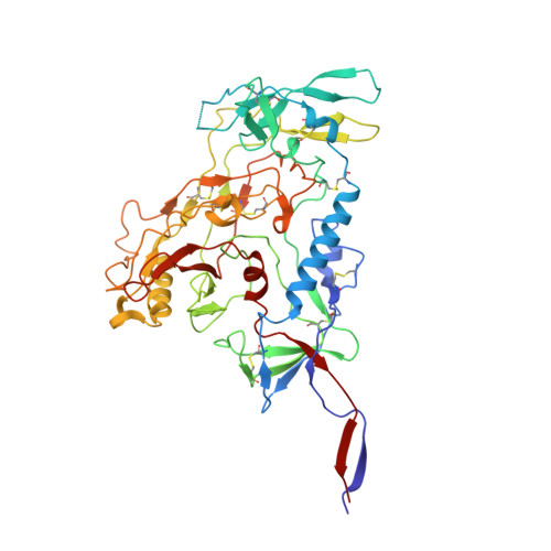
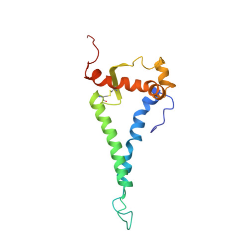
Cryo-ET of Env on intact HIV virions reveals structural variation and positioning on the Gag lattice.

Mangala Prasad, V.Leaman, D.P.Lovendahl, K.N.Croft, J.T.Benhaim, M.A.Hodge, E.A.Zwick, M.B.Lee, K.K.

(2022) Cell 185: 641-653.e17

  • DOI: https://doi.org/10.1016/j.cell.2022.01.013
  • Primary Citation of Related Structures:  
    7SKA

  • PubMed Abstract: 

    HIV-1 Env mediates viral entry into host cells and is the sole target for neutralizing antibodies. However, Env structure and organization in its native virion context has eluded detailed characterization. Here, we used cryo-electron tomography to analyze Env in mature and immature HIV-1 particles. Immature particles showed distinct Env positioning relative to the underlying Gag lattice, providing insights into long-standing questions about Env incorporation. A 9.1-Å sub-tomogram-averaged reconstruction of virion-bound Env in conjunction with structural mass spectrometry revealed unexpected features, including a variable central core of the gp41 subunit, heterogeneous glycosylation between protomers, and a flexible stalk that allows Env tilting and variable exposure of neutralizing epitopes. Together, our results provide an integrative understanding of HIV assembly and structural variation in Env antigen presentation.


  • Organizational Affiliation

    Department of Medicinal Chemistry, University of Washington, Seattle, WA 98195, USA.


Macromolecules
Find similar proteins by:  (by identity cutoff)  |  3D Structure
Entity ID: 1
MoleculeChains Sequence LengthOrganismDetailsImage
Envelope glycoprotein gp120A,
C [auth N],
E [auth Y]
465Human immunodeficiency virus 1Mutation(s): 0 
Gene Names: env
UniProt
Find proteins for Q6TAN8 (Human immunodeficiency virus 1)
Explore Q6TAN8 
Go to UniProtKB:  Q6TAN8
Entity Groups  
Sequence Clusters30% Identity50% Identity70% Identity90% Identity95% Identity100% Identity
UniProt GroupQ6TAN8
Sequence Annotations
Expand
  • Reference Sequence
Find similar proteins by:  (by identity cutoff)  |  3D Structure
Entity ID: 2
MoleculeChains Sequence LengthOrganismDetailsImage
Envelope glycoprotein gp41B,
D [auth O],
F [auth Z]
144Human immunodeficiency virus 1Mutation(s): 0 
Gene Names: env
UniProt
Find proteins for Q6TAN8 (Human immunodeficiency virus 1)
Explore Q6TAN8 
Go to UniProtKB:  Q6TAN8
Entity Groups  
Sequence Clusters30% Identity50% Identity70% Identity90% Identity95% Identity100% Identity
UniProt GroupQ6TAN8
Sequence Annotations
Expand
  • Reference Sequence
Oligosaccharides

Help

Entity ID: 3
MoleculeChains Length2D Diagram Glycosylation3D Interactions
alpha-D-mannopyranose-(1-2)-alpha-D-mannopyranose-(1-3)-beta-D-mannopyranose-(1-4)-2-acetamido-2-deoxy-beta-D-glucopyranose-(1-4)-2-acetamido-2-deoxy-beta-D-glucopyranose
CA [auth b],
G [auth F],
KA [auth j],
N [auth J],
SA [auth s],
CA [auth b],
G [auth F],
KA [auth j],
N [auth J],
SA [auth s],
V [auth T]
5N/AN/A
Entity ID: 4
MoleculeChains Length2D Diagram Glycosylation3D Interactions
alpha-D-mannopyranose-(1-2)-alpha-D-mannopyranose-(1-3)-alpha-D-mannopyranose-(1-6)-beta-D-mannopyranose-(1-4)-2-acetamido-2-deoxy-beta-D-glucopyranose-(1-4)-2-acetamido-2-deoxy-beta-D-glucopyranoseH,
LA [auth k],
W [auth d]
6N/AN/A
Entity ID: 6
MoleculeChains Length2D Diagram Glycosylation3D Interactions
alpha-D-mannopyranose-(1-3)-beta-D-mannopyranose-(1-4)-2-acetamido-2-deoxy-beta-D-glucopyranose-(1-4)-2-acetamido-2-deoxy-beta-D-glucopyranose
BA [auth a],
EA [auth e],
M [auth I],
P [auth L],
RA [auth r],
BA [auth a],
EA [auth e],
M [auth I],
P [auth L],
RA [auth r],
UA [auth u]
4N/AN/A
Entity ID: 7
MoleculeChains Length2D Diagram Glycosylation3D Interactions
beta-D-mannopyranose-(1-4)-2-acetamido-2-deoxy-beta-D-glucopyranose-(1-4)-2-acetamido-2-deoxy-beta-D-glucopyranose
DA [auth c],
FA [auth f],
O [auth K],
Q [auth M],
TA [auth t],
DA [auth c],
FA [auth f],
O [auth K],
Q [auth M],
TA [auth t],
VA [auth v]
3N/AN/A
Entity ID: 8
MoleculeChains Length2D Diagram Glycosylation3D Interactions
2-acetamido-2-deoxy-beta-D-glucopyranose-(1-2)-alpha-D-mannopyranose-(1-3)-beta-D-mannopyranose-(1-4)-2-acetamido-2-deoxy-beta-D-glucopyranose-(1-4)-[alpha-L-fucopyranose-(2-6)]2-acetamido-2-deoxy-beta-D-glucopyranoseGA [auth n],
R [auth P],
WA [auth w]
6N/AN/A
Entity ID: 9
MoleculeChains Length2D Diagram Glycosylation3D Interactions
beta-D-galactopyranose-(1-4)-2-acetamido-2-deoxy-beta-D-glucopyranose-(1-2)-[beta-D-galactopyranose-(1-4)-2-acetamido-2-deoxy-beta-D-glucopyranose-(1-4)]alpha-D-mannopyranose-(1-3)-beta-D-mannopyranose-(1-4)-2-acetamido-2-deoxy-beta-D-glucopyranose-(1-4)-2-acetamido-2-deoxy-beta-D-glucopyranoseHA [auth g],
S [auth Q],
XA [auth x]
8N/AN/A
Entity ID: 10
MoleculeChains Length2D Diagram Glycosylation3D Interactions
2-acetamido-2-deoxy-beta-D-glucopyranose-(1-2)-alpha-D-mannopyranoseIA [auth h],
T [auth R],
YA [auth y]
2N/AN/A
Entity ID: 11
MoleculeChains Length2D Diagram Glycosylation3D Interactions
alpha-D-mannopyranose-(1-3)-alpha-D-mannopyranose-(1-6)-[alpha-D-mannopyranose-(1-3)]beta-D-mannopyranose-(1-4)-2-acetamido-2-deoxy-beta-D-glucopyranose-(1-4)-2-acetamido-2-deoxy-beta-D-glucopyranoseJA [auth i],
NA [auth m],
U [auth S]
6N/AN/A
Small Molecules
Ligands 2 Unique
IDChains Name / Formula / InChI Key2D Diagram3D Interactions
NAG
Query on NAG

Download Ideal Coordinates CCD File 
AB [auth A]
BB [auth A]
CB [auth A]
DB [auth A]
EB [auth B]
AB [auth A],
BB [auth A],
CB [auth A],
DB [auth A],
EB [auth B],
FB [auth B],
HB [auth N],
IB [auth N],
JB [auth N],
KB [auth N],
LB [auth N],
MB [auth O],
NB [auth O],
PB [auth Y],
QB [auth Y],
RB [auth Y],
SB [auth Y],
TB [auth Y],
UB [auth Z],
VB [auth Z],
ZA [auth A]
2-acetamido-2-deoxy-beta-D-glucopyranose
C8 H15 N O6
OVRNDRQMDRJTHS-FMDGEEDCSA-N
FUC
Query on FUC

Download Ideal Coordinates CCD File 
GB [auth B],
OB [auth O],
WB [auth Z]
alpha-L-fucopyranose
C6 H12 O5
SHZGCJCMOBCMKK-SXUWKVJYSA-N
Experimental Data & Validation

Experimental Data

  • Method: ELECTRON MICROSCOPY
  • Resolution: 9.10 Å
  • Aggregation State: PARTICLE 
  • Reconstruction Method: SUBTOMOGRAM AVERAGING 
EM Software:
TaskSoftware PackageVersion
RECONSTRUCTIONEMAN2
MODEL REFINEMENTCoot

Structure Validation

View Full Validation Report


Entry History & Funding Information

Deposition Data


Funding OrganizationLocationGrant Number
National Institutes of Health/National Institute Of Allergy and Infectious Diseases (NIH/NIAID)United StatesR01AI140868
National Institutes of Health/National Institute of General Medical Sciences (NIH/NIGMS)United StatesR01GM099989
National Institutes of Health/National Institute Of Allergy and Infectious Diseases (NIH/NIAID)United StatesR01AI143563

Revision History  (Full details and data files)

  • Version 1.0: 2022-03-09
    Type: Initial release