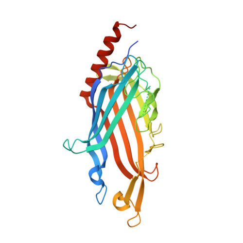
Crystal structure of the putative cell-wall lipoglycan biosynthesis protein LmcA from Mycobacterium smegmatis.
Patel, O., Brammananth, R., Dai, W., Panjikar, S., Coppel, R.L., Lucet, I.S., Crellin, P.K.(2022) Acta Crystallogr D Struct Biol 78: 494-508
- PubMed: 35362472 Search on PubMedSearch on PubMed Central
- DOI: https://doi.org/10.1107/S2059798322001772
- Primary Citation Related Structures:
7N3V, 7SHW - PubMed Abstract:
The bacterial genus Mycobacterium includes important pathogens, most notably M. tuberculosis, which infects one-quarter of the entire human population, resulting in around 1.4 million deaths from tuberculosis each year. Mycobacteria, and the closely related corynebacteria, synthesize a class of abundant glycolipids, the phosphatidyl-myo-inositol mannosides (PIMs). PIMs serve as membrane anchors for hyperglycosylated species, lipomannan (LM) and lipoarabinomannan (LAM), which are surface-exposed and modulate the host immune response. Previously, in studies using the model species Corynebacterium glutamicum, NCgl2760 was identified as a novel membrane protein that is required for the synthesis of full-length LM and LAM. Here, the first crystal structure of its ortholog in Mycobacterium smegmatis, MSMEG_0317, is reported at 1.8 Å resolution. The structure revealed an elongated β-barrel fold enclosing two distinct cavities and one α-helix extending away from the β-barrel core, resembling a `cone with a flake' arrangement. Through xenon derivatization and structural comparison with AlphaFold2-derived predictions of the M. tuberculosis homolog Rv0227c, structural elements were identified that may undergo conformational changes to switch from `closed' to `open' conformations, allowing cavity access. An AlphaFold2-derived NCgl2760 model predicted a smaller β-barrel core with an enclosed central cavity, suggesting that all three proteins, which were collectively termed LmcA, may have a common mechanism of ligand binding through these cavities. These findings provide new structural insights into the biosynthetic pathway for a family of surface lipoglycans with important roles in mycobacterial pathogenesis.
- The Walter and Eliza Hall Institute of Medical Research, Parkville, Victoria 3052, Australia.
Organizational Affiliation: