7SHF

Cryo-EM structure of GPR158 coupled to the RGS7-Gbeta5 complex


Experimental Data Snapshot

  • Method: ELECTRON MICROSCOPY
  • Resolution: 3.40 Å
  • Aggregation State: PARTICLE 
  • Reconstruction Method: SINGLE PARTICLE 

wwPDB Validation   3D Report Full Report


This is version 2.1 of the entry. See complete history


Literature

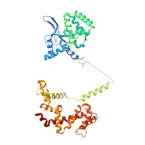
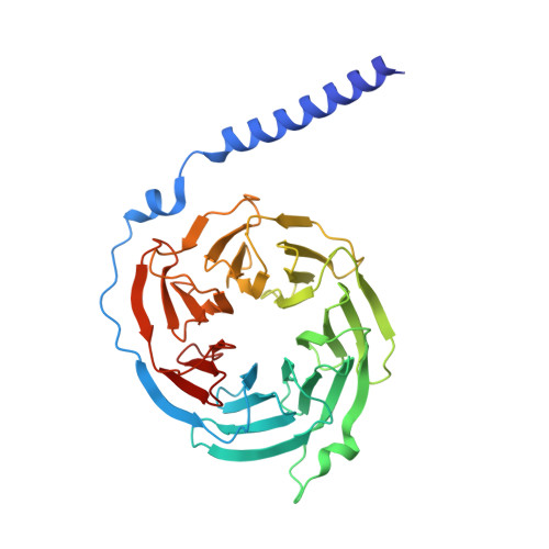
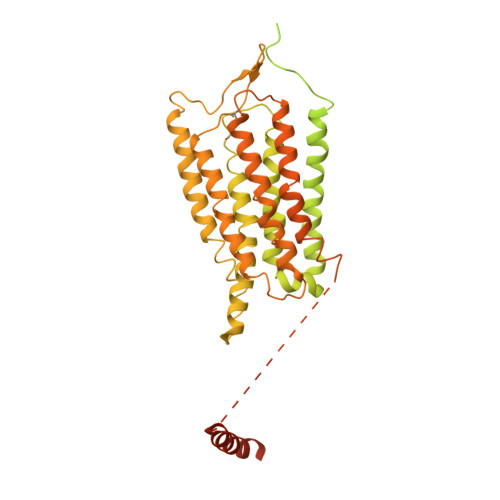
Cryo-EM structure of human GPR158 receptor coupled to the RGS7-G beta 5 signaling complex.

Patil, D.N.Singh, S.Laboute, T.Strutzenberg, T.S.Qiu, X.Wu, D.Novick, S.J.Robinson, C.V.Griffin, P.R.Hunt, J.F.Izard, T.Singh, A.K.Martemyanov, K.A.

(2022) Science 375: 86-91

  • DOI: https://doi.org/10.1126/science.abl4732
  • Primary Citation of Related Structures:  
    7SHE, 7SHF

  • PubMed Abstract: 

    GPR158 is an orphan G protein–coupled receptor (GPCR) highly expressed in the brain, where it controls synapse formation and function. GPR158 has also been implicated in depression, carcinogenesis, and cognition. However, the structural organization and signaling mechanisms of GPR158 are largely unknown. We used single-particle cryo–electron microscopy (cryo-EM) to determine the structures of human GPR158 alone and bound to an RGS signaling complex. The structures reveal a homodimeric organization stabilized by a pair of phospholipids and the presence of an extracellular Cache domain, an unusual ligand-binding domain in GPCRs. We further demonstrate the structural basis of GPR158 coupling to RGS7-Gβ5. Together, these results provide insights into the unusual biology of orphan receptors and the formation of GPCR-RGS complexes.


  • Organizational Affiliation

    Department of Neuroscience, The Scripps Research Institute, Jupiter, FL 33458, USA.


Macromolecules
Find similar proteins by:  (by identity cutoff)  |  3D Structure
Entity ID: 1
MoleculeChains Sequence LengthOrganismDetailsImage
Isoform 2 of Regulator of G-protein signaling 7A [auth C]469Homo sapiensMutation(s): 0 
Gene Names: RGS7
Membrane Entity: Yes 
UniProt & NIH Common Fund Data Resources
Find proteins for P49802 (Homo sapiens)
Explore P49802 
Go to UniProtKB:  P49802
PHAROS:  P49802
GTEx:  ENSG00000182901 
Entity Groups  
Sequence Clusters30% Identity50% Identity70% Identity90% Identity95% Identity100% Identity
UniProt GroupP49802
Sequence Annotations
Expand
  • Reference Sequence
Find similar proteins by:  (by identity cutoff)  |  3D Structure
Entity ID: 2
MoleculeChains Sequence LengthOrganismDetailsImage
Guanine nucleotide-binding protein subunit beta-5B [auth D]353Mus musculusMutation(s): 0 
Gene Names: Gnb5
Membrane Entity: Yes 
UniProt & NIH Common Fund Data Resources
Find proteins for P62881 (Mus musculus)
Explore P62881 
Go to UniProtKB:  P62881
IMPC:  MGI:101848
Entity Groups  
Sequence Clusters30% Identity50% Identity70% Identity90% Identity95% Identity100% Identity
UniProt GroupP62881
Sequence Annotations
Expand
  • Reference Sequence
Find similar proteins by:  (by identity cutoff)  |  3D Structure
Entity ID: 3
MoleculeChains Sequence LengthOrganismDetailsImage
G-protein coupled receptor 158C [auth B],
D [auth A]
781Homo sapiensMutation(s): 0 
Gene Names: GPR158KIAA1136
Membrane Entity: Yes 
UniProt & NIH Common Fund Data Resources
Find proteins for Q5T848 (Homo sapiens)
Explore Q5T848 
Go to UniProtKB:  Q5T848
PHAROS:  Q5T848
GTEx:  ENSG00000151025 
Entity Groups  
Sequence Clusters30% Identity50% Identity70% Identity90% Identity95% Identity100% Identity
UniProt GroupQ5T848
Sequence Annotations
Expand
  • Reference Sequence
Small Molecules
Ligands 3 Unique
IDChains Name / Formula / InChI Key2D Diagram3D Interactions
EIJ
Query on EIJ

Download Ideal Coordinates CCD File 
AA [auth A](2S)-1-{[(S)-hydroxy{[(1s,2R,3R,4R,5S,6S)-2,3,4,5,6-pentahydroxycyclohexyl]oxy}phosphoryl]oxy}-3-(octadecanoyloxy)propan-2-yl (5E,8E,11E,14E)-icosa-5,8,11,14-tetraenoate
C47 H83 O13 P
KRTOMQDUKGRFDJ-ROVRCULJSA-N
PEE
Query on PEE

Download Ideal Coordinates CCD File 
BA [auth A]1,2-dioleoyl-sn-glycero-3-phosphoethanolamine
C41 H78 N O8 P
MWRBNPKJOOWZPW-NYVOMTAGSA-N
CLR
Query on CLR

Download Ideal Coordinates CCD File 
E [auth B]
F [auth B]
G [auth B]
H [auth B]
I [auth B]
E [auth B],
F [auth B],
G [auth B],
H [auth B],
I [auth B],
J [auth B],
K [auth B],
L [auth B],
M [auth B],
N [auth B],
O [auth A],
P [auth A],
Q [auth A],
R [auth A],
S [auth A],
T [auth A],
U [auth A],
V [auth A],
W [auth A],
X [auth A],
Y [auth A],
Z [auth A]
CHOLESTEROL
C27 H46 O
HVYWMOMLDIMFJA-DPAQBDIFSA-N
Experimental Data & Validation

Experimental Data

  • Method: ELECTRON MICROSCOPY
  • Resolution: 3.40 Å
  • Aggregation State: PARTICLE 
  • Reconstruction Method: SINGLE PARTICLE 
EM Software:
TaskSoftware PackageVersion
RECONSTRUCTIONcryoSPARC

Structure Validation

View Full Validation Report


Entry History & Funding Information

Deposition Data


Funding OrganizationLocationGrant Number
National Institutes of Health/National Institute of Mental Health (NIH/NIMH)United States--

Revision History  (Full details and data files)

  • Version 1.0: 2021-12-01
    Type: Initial release
  • Version 2.0: 2021-12-29
    Type: Coordinate replacement
    Reason: Atomic clashes
    Changes: Advisory, Atomic model, Author supporting evidence, Data collection, Data processing, Database references, Derived calculations, Non-polymer description, Polymer sequence, Source and taxonomy, Structure summary
  • Version 2.1: 2022-01-19
    Changes: Database references