7SH1

Class II UvrA protein - Ecm16


Experimental Data Snapshot

  • Method: X-RAY DIFFRACTION
  • Resolution: 2.04 Å
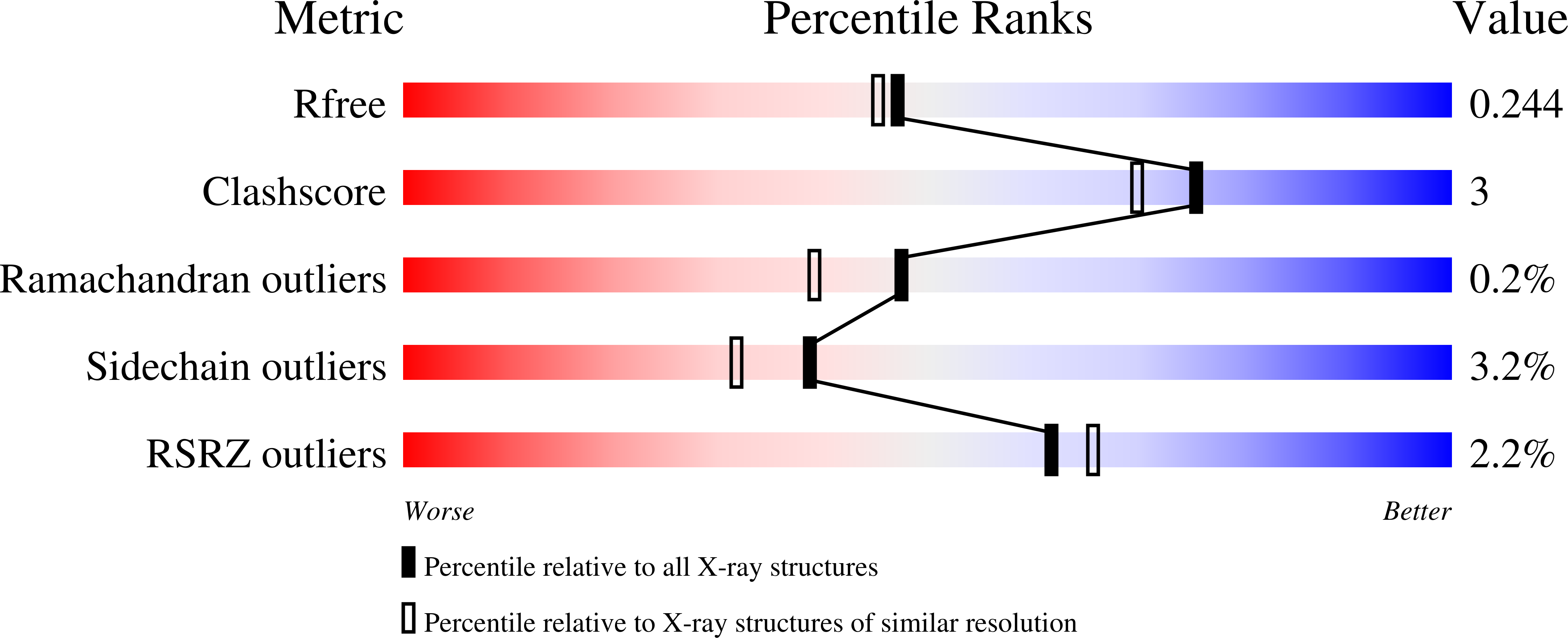
  • R-Value Free: 0.239 
  • R-Value Work: 0.198 
  • R-Value Observed: 0.200 

Starting Models: experimental
View more details

wwPDB Validation   3D Report Full Report


Ligand Structure Quality Assessment 


This is version 2.2 of the entry. See complete history


Literature

Structural and functional analyses of the echinomycin resistance conferring protein Ecm16 from Streptomyces lasalocidi.

Gade, P.Erlandson, A.Ullah, A.Chen, X.Mathews, I.I.Mera, P.E.Kim, C.Y.

(2023) Sci Rep 13: 7980-7980

  • DOI: https://doi.org/10.1038/s41598-023-34437-9
  • Primary Citation of Related Structures:  
    7SH1

  • PubMed Abstract: 

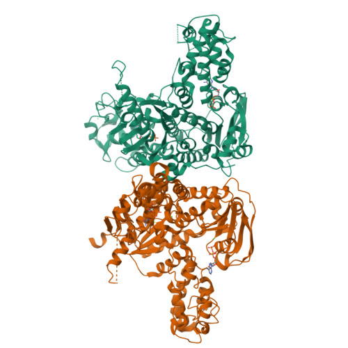
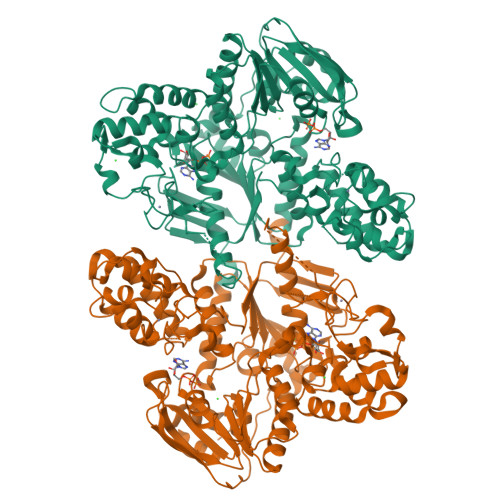
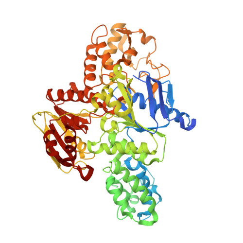
    Echinomycin is a natural product DNA bisintercalator antibiotic. The echinomycin biosynthetic gene cluster in Streptomyces lasalocidi includes a gene encoding the self-resistance protein Ecm16. Here, we present the 2.0 Å resolution crystal structure of Ecm16 bound to adenosine diphosphate. The structure of Ecm16 closely resembles that of UvrA, the DNA damage sensor component of the prokaryotic nucleotide excision repair system, but Ecm16 lacks the UvrB-binding domain and its associated zinc-binding module found in UvrA. Mutagenesis study revealed that the insertion domain of Ecm16 is required for DNA binding. Furthermore, the specific amino acid sequence of the insertion domain allows Ecm16 to distinguish echinomycin-bound DNA from normal DNA and link substrate binding to ATP hydrolysis activity. Expression of ecm16 in the heterologous host Brevibacillus choshinensis conferred resistance against echinomycin and other quinomycin antibiotics, including thiocoraline, quinaldopeptin, and sandramycin. Our study provides new insight into how the producers of DNA bisintercalator antibiotics fend off the toxic compounds that they produce.


  • Organizational Affiliation

    Department of Chemistry and Biochemistry, The University of Texas at El Paso, El Paso, TX, USA.


Macromolecules
Find similar proteins by:  (by identity cutoff)  |  3D Structure
Entity ID: 1
MoleculeChains Sequence LengthOrganismDetailsImage
Excinuclease ABC subunit UvrA
A, B
812Streptomyces lasalocidiMutation(s): 0 
Gene Names: ecm16E4U91_36965
UniProt
Find proteins for Q0X0A9 (Streptomyces lasalocidi)
Explore Q0X0A9 
Go to UniProtKB:  Q0X0A9
Entity Groups  
Sequence Clusters30% Identity50% Identity70% Identity90% Identity95% Identity100% Identity
UniProt GroupQ0X0A9
Sequence Annotations
Expand
  • Reference Sequence
Small Molecules
Ligands 4 Unique
IDChains Name / Formula / InChI Key2D Diagram3D Interactions
ADP (Subject of Investigation/LOI)
Query on ADP

Download Ideal Coordinates CCD File 
C [auth A],
D [auth A],
K [auth B],
L [auth B]
ADENOSINE-5'-DIPHOSPHATE
C10 H15 N5 O10 P2
XTWYTFMLZFPYCI-KQYNXXCUSA-N
ZN (Subject of Investigation/LOI)
Query on ZN

Download Ideal Coordinates CCD File 
E [auth A],
F [auth A],
M [auth B],
N [auth B]
ZINC ION
Zn
PTFCDOFLOPIGGS-UHFFFAOYSA-N
CL (Subject of Investigation/LOI)
Query on CL

Download Ideal Coordinates CCD File 
G [auth A],
H [auth A],
I [auth A],
O [auth B],
P [auth B]
CHLORIDE ION
Cl
VEXZGXHMUGYJMC-UHFFFAOYSA-M
MG (Subject of Investigation/LOI)
Query on MG

Download Ideal Coordinates CCD File 
J [auth A],
Q [auth B]
MAGNESIUM ION
Mg
JLVVSXFLKOJNIY-UHFFFAOYSA-N
Experimental Data & Validation

Experimental Data

  • Method: X-RAY DIFFRACTION
  • Resolution: 2.04 Å
  • R-Value Free: 0.239 
  • R-Value Work: 0.198 
  • R-Value Observed: 0.200 
  • Space Group: P 31 2 1
Unit Cell:
Length ( Å )Angle ( ˚ )
a = 141.13α = 90
b = 141.13β = 90
c = 173.614γ = 120
Software Package:
Software NamePurpose
REFMACrefinement
XDSdata reduction
XDSdata scaling
PHASERphasing

Structure Validation

View Full Validation Report


Ligand Structure Quality Assessment 


Entry History & Funding Information

Deposition Data


Funding OrganizationLocationGrant Number
Other governmentUnited States--

Revision History  (Full details and data files)

  • Version 1.0: 2022-10-26
    Type: Initial release
  • Version 2.0: 2023-05-24
    Type: Coordinate replacement
    Reason: Ligand geometry
    Changes: Advisory, Atomic model, Author supporting evidence, Data collection, Database references, Derived calculations, Non-polymer description, Other, Refinement description, Source and taxonomy, Structure summary
  • Version 2.1: 2023-05-31
    Changes: Database references
  • Version 2.2: 2023-10-25
    Changes: Data collection, Refinement description