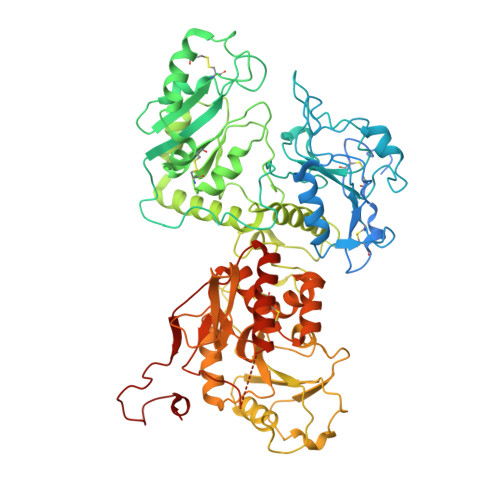
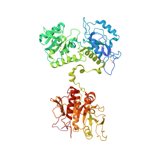
7SCK

Cryo-EM structure of the human Exostosin-1 and Exostosin-2 heterodimer in complex with a 7-sugar oligosaccharide acceptor analog

  • Classification: TRANSFERASE
  • Organism(s): Homo sapiens
  • Expression System: Homo sapiens
  • Mutation(s): No 

  • Deposited: 2021-09-28 Released: 2022-09-28 
  • Deposition Author(s): Li, H., Li, H.
  • Funding Organization(s): National Institutes of Health/National Cancer Institute (NIH/NCI)

Experimental Data Snapshot

  • Method: ELECTRON MICROSCOPY
  • Resolution: 2.80 Å
  • Aggregation State: PARTICLE 
  • Reconstruction Method: SINGLE PARTICLE 

wwPDB Validation   3D Report Full Report


This is version 1.2 of the entry. See complete history


Literature

Structural basis for heparan sulfate co-polymerase action by the EXT1-2 complex.

Li, H.Chapla, D.Amos, R.A.Ramiah, A.Moremen, K.W.Li, H.

(2023) Nat Chem Biol 19: 565-574

  • DOI: https://doi.org/10.1038/s41589-022-01220-2
  • Primary Citation of Related Structures:  
    7SCH, 7SCJ, 7SCK, 7UQX, 7UQY

  • PubMed Abstract: 

    Heparan sulfate (HS) proteoglycans are extended (-GlcAβ1,4GlcNAcα1,4-) n co-polymers containing decorations of sulfation and epimerization that are linked to cell surface and extracellular matrix proteins. In mammals, HS repeat units are extended by an obligate heterocomplex of two exostosin family members, EXT1 and EXT2, where each protein monomer contains distinct GT47 (GT-B fold) and GT64 (GT-A fold) glycosyltransferase domains. In this study, we generated human EXT1-EXT2 (EXT1-2) as a functional heterocomplex and determined its structure in the presence of bound donor and acceptor substrates. Structural data and enzyme activity of catalytic site mutants demonstrate that only two of the four glycosyltransferase domains are major contributors to co-polymer syntheses: the EXT1 GT-B fold β1,4GlcA transferase domain and the EXT2 GT-A fold α1,4GlcNAc transferase domain. The two catalytic sites are over 90 Å apart, indicating that HS is synthesized by a dissociative process that involves a single catalytic site on each monomer.


  • Organizational Affiliation

    Department of Structural Biology, Van Andel Institute, Grand Rapids, MI, USA.


Macromolecules
Find similar proteins by:  (by identity cutoff)  |  3D Structure
Entity ID: 1
MoleculeChains Sequence LengthOrganismDetailsImage
Exostosin-1720Homo sapiensMutation(s): 0 
Gene Names: EXT1
EC: 2.4.1.224 (PDB Primary Data), 2.4.1.225 (PDB Primary Data)
UniProt & NIH Common Fund Data Resources
Find proteins for Q16394 (Homo sapiens)
Explore Q16394 
Go to UniProtKB:  Q16394
PHAROS:  Q16394
GTEx:  ENSG00000182197 
Entity Groups  
Sequence Clusters30% Identity50% Identity70% Identity90% Identity95% Identity100% Identity
UniProt GroupQ16394
Glycosylation
Glycosylation Sites: 1Go to GlyGen: Q16394-1
Sequence Annotations
Expand
  • Reference Sequence
Find similar proteins by:  (by identity cutoff)  |  3D Structure
Entity ID: 2
MoleculeChains Sequence LengthOrganismDetailsImage
Exostosin-2674Homo sapiensMutation(s): 0 
Gene Names: EXT2
EC: 2.4.1.224 (PDB Primary Data), 2.4.1.225 (PDB Primary Data)
UniProt & NIH Common Fund Data Resources
Find proteins for Q93063 (Homo sapiens)
Explore Q93063 
Go to UniProtKB:  Q93063
PHAROS:  Q93063
GTEx:  ENSG00000151348 
Entity Groups  
Sequence Clusters30% Identity50% Identity70% Identity90% Identity95% Identity100% Identity
UniProt GroupQ93063
Glycosylation
Glycosylation Sites: 1Go to GlyGen: Q93063-1
Sequence Annotations
Expand
  • Reference Sequence
Oligosaccharides

Help

Entity ID: 3
MoleculeChains Length2D Diagram Glycosylation3D Interactions
beta-D-mannopyranose-(1-4)-2-acetamido-2-deoxy-beta-D-glucopyranose-(1-4)-2-acetamido-2-deoxy-beta-D-glucopyranose
C
3N/AN-Glycosylation
Glycosylation Resources
GlyTouCan:  G15407YE
GlyCosmos:  G15407YE
GlyGen:  G15407YE
Entity ID: 4
MoleculeChains Length2D Diagram Glycosylation3D Interactions
beta-D-glucopyranuronic acid-(1-4)-2-acetamido-2-deoxy-alpha-D-glucopyranose-(1-4)-beta-D-glucopyranuronic acid-(1-4)-2-acetamido-2-deoxy-alpha-D-glucopyranose
D
4N/AN/A
Experimental Data & Validation

Experimental Data

  • Method: ELECTRON MICROSCOPY
  • Resolution: 2.80 Å
  • Aggregation State: PARTICLE 
  • Reconstruction Method: SINGLE PARTICLE 
EM Software:
TaskSoftware PackageVersion
RECONSTRUCTIONRELION3.1

Structure Validation

View Full Validation Report


Entry History & Funding Information

Deposition Data

  • Released Date: 2022-09-28 
  • Deposition Author(s): Li, H., Li, H.

Funding OrganizationLocationGrant Number
National Institutes of Health/National Cancer Institute (NIH/NCI)United StatesCA231466

Revision History  (Full details and data files)

  • Version 1.0: 2022-09-28
    Type: Initial release
  • Version 1.1: 2023-01-18
    Changes: Database references
  • Version 1.2: 2023-05-17
    Changes: Database references