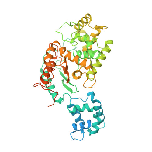
7SBM

Human glutaminase C (Y466W) with L-Gln, open conformation

  • Classification: HYDROLASE
  • Organism(s): Homo sapiens
  • Expression System: Escherichia coli
  • Mutation(s): Yes 

  • Deposited: 2021-09-25 Released: 2022-02-02 
  • Deposition Author(s): Nguyen, T.-T.T., Cerione, R.A.
  • Funding Organization(s): National Institutes of Health/National Cancer Institute (NIH/NCI), National Institutes of Health/National Institute of General Medical Sciences (NIH/NIGMS)

Experimental Data Snapshot

  • Method: X-RAY DIFFRACTION
  • Resolution: 2.80 Å
  • R-Value Free: 0.243 
  • R-Value Work: 0.200 
  • R-Value Observed: 0.202 

Starting Model: experimental
View more details

wwPDB Validation   3D Report Full Report


Ligand Structure Quality Assessment 


This is version 1.2 of the entry. See complete history


Literature

High-resolution structures of mitochondrial glutaminase C tetramers indicate conformational changes upon phosphate binding.

Nguyen, T.T.Ramachandran, S.Hill, M.J.Cerione, R.A.

(2022) J Biol Chem 298: 101564-101564

  • DOI: https://doi.org/10.1016/j.jbc.2022.101564
  • Primary Citation of Related Structures:  
    7SBM, 7SBN

  • PubMed Abstract: 

    The mitochondrial enzyme glutaminase C (GAC) is upregulated in many cancer cells to catalyze the first step in glutamine metabolism, the hydrolysis of glutamine to glutamate. The dependence of cancer cells on this transformed metabolic pathway highlights GAC as a potentially important therapeutic target. GAC acquires maximal catalytic activity upon binding to anionic activators such as inorganic phosphate. To delineate the mechanism of GAC activation, we used the tryptophan substitution of tyrosine 466 in the catalytic site of the enzyme as a fluorescent reporter for glutamine binding in the presence and absence of phosphate. We show that in the absence of phosphate, glutamine binding to the Y466W GAC tetramer exhibits positive cooperativity. A high-resolution X-ray structure of tetrameric Y466W GAC bound to glutamine suggests that cooperativity in substrate binding is coupled to tyrosine 249, located at the edge of the catalytic site (i.e., the "lid"), adopting two distinct conformations. In one dimer within the GAC tetramer, the lids are open and glutamine binds weakly, whereas, in the adjoining dimer, the lids are closed over the substrates, resulting in higher affinity interactions. When crystallized in the presence of glutamine and phosphate, all four subunits of the Y466W GAC tetramer exhibited bound glutamine with closed lids. Glutamine can bind with high affinity to each subunit, which subsequently undergo simultaneous catalysis. These findings explain how the regulated transitioning of GAC between different conformational states ensures that maximal catalytic activity is reached in cancer cells only when an allosteric activator is available.


  • Organizational Affiliation

    Department of Chemistry and Chemical Biology, Cornell University, Ithaca, New York, USA.


Macromolecules
Find similar proteins by:  (by identity cutoff)  |  3D Structure
Entity ID: 1
MoleculeChains Sequence LengthOrganismDetailsImage
Isoform 3 of Glutaminase kidney isoform, mitochondrial
A, B, C, D
539Homo sapiensMutation(s): 1 
Gene Names: GLSGLS1KIAA0838
EC: 3.5.1.2
UniProt & NIH Common Fund Data Resources
Find proteins for O94925 (Homo sapiens)
Explore O94925 
Go to UniProtKB:  O94925
PHAROS:  O94925
GTEx:  ENSG00000115419 
Entity Groups  
Sequence Clusters30% Identity50% Identity70% Identity90% Identity95% Identity100% Identity
UniProt GroupO94925
Sequence Annotations
Expand
  • Reference Sequence
Experimental Data & Validation

Experimental Data

  • Method: X-RAY DIFFRACTION
  • Resolution: 2.80 Å
  • R-Value Free: 0.243 
  • R-Value Work: 0.200 
  • R-Value Observed: 0.202 
  • Space Group: P 21 21 21
Unit Cell:
Length ( Å )Angle ( ˚ )
a = 98.842α = 90
b = 139.1β = 90
c = 177.435γ = 90
Software Package:
Software NamePurpose
PHENIXrefinement
HKL-2000data reduction
HKL-2000data scaling
PHENIXphasing

Structure Validation

View Full Validation Report


Ligand Structure Quality Assessment 


Entry History & Funding Information

Deposition Data


Funding OrganizationLocationGrant Number
National Institutes of Health/National Cancer Institute (NIH/NCI)United StatesCA201402
National Institutes of Health/National Institute of General Medical Sciences (NIH/NIGMS)United StatesGM122575

Revision History  (Full details and data files)

  • Version 1.0: 2022-02-02
    Type: Initial release
  • Version 1.1: 2022-02-09
    Changes: Database references
  • Version 1.2: 2023-10-18
    Changes: Data collection, Refinement description