7SB8 | pdb_00007sb8

d(GA(CGA)5) parallel-stranded homo-duplex


Experimental Data Snapshot

  • Method: X-RAY DIFFRACTION
  • Resolution: 1.32 Å
  • R-Value Free: 
    0.214 (Depositor), 0.214 (DCC) 
  • R-Value Work: 
    0.171 (Depositor), 0.172 (DCC) 
  • R-Value Observed: 
    0.173 (Depositor) 

Starting Model: experimental
View more details

wwPDB Validation 3D Report Full Report

Validation slider image for 7SB8

This is version 1.2 of the entry. See complete history

Literature

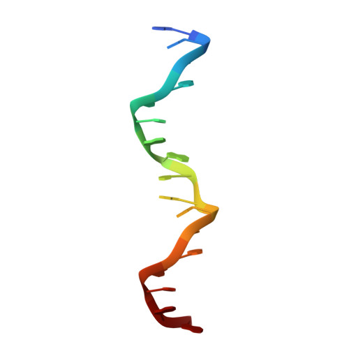
The parallel-stranded d(CGA) duplex is a highly predictable structural motif with two conformationally distinct strands.

Luteran, E.M.Paukstelis, P.J.

(2022) Acta Crystallogr D Struct Biol 78: 299-309

  • DOI: https://doi.org/10.1107/S2059798322000304
  • Primary Citation Related Structures: 
    7SB8, 7T6Y

  • PubMed Abstract: 

    DNA can adopt noncanonical structures that have important biological functions while also providing structural diversity for applications in nanotechnology. Here, the crystal structures of two oligonucleotides composed of d(CGA) triplet repeats in the parallel-stranded duplex form are described. The structure determination of four unique d(CGA)-based parallel-stranded duplexes across two crystal structures has allowed the structural parameters of d(CGA) triplets in the parallel-stranded duplex form to be characterized and established. These results show that d(CGA) units are highly uniform, but that each strand in the duplex is structurally unique and has a distinct role in accommodating structural asymmetries induced by the C-CH + base pair.


  • Organizational Affiliation
    • Department of Chemistry and Biochemistry, University of Maryland, College Park, MD 20742, USA.

Macromolecule Content 

  • Total Structure Weight: 33.66 kDa 
  • Atom Count: 2,680 
  • Modeled Residue Count: 102 
  • Deposited Residue Count: 102 
  • Unique nucleic acid chains: 1

Macromolecules

Find similar nucleic acids by:  Sequence
Entity ID: 1
MoleculeChains LengthOrganismImage
GA(CGA)517synthetic construct
Sequence Annotations
Expand
Reference Sequence

Small Molecules

Ligands 3 Unique
IDChains Name / Formula / InChI Key2D Diagram3D Interactions
NCO

Query on NCO


Download:Ideal Coordinates CCD File
M [auth B]
N [auth B]
Q [auth D]
R [auth D]
S [auth D]
M [auth B],
N [auth B],
Q [auth D],
R [auth D],
S [auth D],
X [auth F]
COBALT HEXAMMINE(III)
Co H18 N6
DYLMFCCYOUSRTK-UHFFFAOYSA-N
SR

Query on SR


Download:Ideal Coordinates CCD File
G [auth A]
H [auth A]
I [auth A]
J [auth B]
K [auth B]
G [auth A],
H [auth A],
I [auth A],
J [auth B],
K [auth B],
O [auth D],
P [auth D],
T [auth E],
U [auth E],
V [auth F],
W [auth F],
Y [auth C],
Z [auth C]
STRONTIUM ION
Sr
PWYYWQHXAPXYMF-UHFFFAOYSA-N
NA

Query on NA


Download:Ideal Coordinates CCD File
L [auth B]SODIUM ION
Na
FKNQFGJONOIPTF-UHFFFAOYSA-N

Experimental Data & Validation

Experimental Data

  • Method: X-RAY DIFFRACTION
  • Resolution: 1.32 Å
  • R-Value Free:  0.214 (Depositor), 0.214 (DCC) 
  • R-Value Work:  0.171 (Depositor), 0.172 (DCC) 
  • R-Value Observed: 0.173 (Depositor) 
Space Group: P 1 21 1
Unit Cell:
Length ( Å )Angle ( ˚ )
a = 19.677α = 90
b = 30.422β = 90.41
c = 180.822γ = 90
Software Package:
Software NamePurpose
PHENIXrefinement
PHASERphasing
PDB_EXTRACTdata extraction
XDSdata reduction
Aimlessdata scaling

Structure Validation

View Full Validation Report


Entry History 

& Funding Information

Deposition Data


Funding OrganizationLocationGrant Number
Not funded--

Revision History  (Full details and data files)

  • Version 1.0: 2021-10-06
    Type: Initial release
  • Version 1.1: 2022-03-16
    Changes: Database references
  • Version 1.2: 2023-10-18
    Changes: Data collection, Refinement description