7SAQ

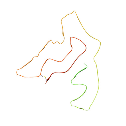
Cryo-EM structure of TMEM106B fibrils extracted from a FTLD-TDP patient, polymorph 1


Experimental Data Snapshot

  • Method: ELECTRON MICROSCOPY
  • Resolution: 2.90 Å
  • Aggregation State: FILAMENT 
  • Reconstruction Method: HELICAL 

wwPDB Validation   3D Report Full Report


This is version 1.3 of the entry. See complete history


Literature

Amyloid fibrils in FTLD-TDP are composed of TMEM106B and not TDP-43.

Jiang, Y.X.Cao, Q.Sawaya, M.R.Abskharon, R.Ge, P.DeTure, M.Dickson, D.W.Fu, J.Y.Ogorzalek Loo, R.R.Loo, J.A.Eisenberg, D.S.

(2022) Nature 605: 304-309

  • DOI: https://doi.org/10.1038/s41586-022-04670-9
  • Primary Citation of Related Structures:  
    7SAQ, 7SAR, 7SAS

  • PubMed Abstract: 

    Frontotemporal lobar degeneration (FTLD) is the third most common neurodegenerative condition after Alzheimer's and Parkinson's diseases 1 . FTLD typically presents in 45 to 64 year olds with behavioural changes or progressive decline of language skills 2 . The subtype FTLD-TDP is characterized by certain clinical symptoms and pathological neuronal inclusions with TAR DNA-binding protein (TDP-43) immunoreactivity 3 . Here we extracted amyloid fibrils from brains of four patients representing four of the five FTLD-TDP subclasses, and determined their structures by cryo-electron microscopy. Unexpectedly, all amyloid fibrils examined were composed of a 135-residue carboxy-terminal fragment of transmembrane protein 106B (TMEM106B), a lysosomal membrane protein previously implicated as a genetic risk factor for FTLD-TDP 4 . In addition to TMEM106B fibrils, we detected abundant non-fibrillar aggregated TDP-43 by immunogold labelling. Our observations confirm that FTLD-TDP is associated with amyloid fibrils, and that the fibrils are formed by TMEM106B rather than TDP-43.


  • Organizational Affiliation

    Departments of Chemistry and Biochemistry and Biological Chemistry, UCLA-DOE Institute, and Molecular Biology Institute, UCLA, Los Angeles, CA, USA.


Macromolecules
Find similar proteins by:  (by identity cutoff)  |  3D Structure
Entity ID: 1
MoleculeChains Sequence LengthOrganismDetailsImage
Transmembrane protein 106B
A, B, C, D, E
274Homo sapiensMutation(s): 0 
UniProt & NIH Common Fund Data Resources
Find proteins for Q9NUM4 (Homo sapiens)
Explore Q9NUM4 
Go to UniProtKB:  Q9NUM4
PHAROS:  Q9NUM4
GTEx:  ENSG00000106460 
Entity Groups  
Sequence Clusters30% Identity50% Identity70% Identity90% Identity95% Identity100% Identity
UniProt GroupQ9NUM4
Sequence Annotations
Expand
  • Reference Sequence
Small Molecules
Ligands 1 Unique
IDChains Name / Formula / InChI Key2D Diagram3D Interactions
NAG
Query on NAG

Download Ideal Coordinates CCD File 
F [auth A]
G [auth A]
H [auth A]
I [auth A]
J [auth B]
F [auth A],
G [auth A],
H [auth A],
I [auth A],
J [auth B],
K [auth B],
L [auth B],
M [auth B],
N [auth C],
O [auth C],
P [auth C],
Q [auth C],
R [auth D],
S [auth D],
T [auth D],
U [auth D],
V [auth E],
W [auth E],
X [auth E],
Y [auth E]
2-acetamido-2-deoxy-beta-D-glucopyranose
C8 H15 N O6
OVRNDRQMDRJTHS-FMDGEEDCSA-N
Experimental Data & Validation

Experimental Data

  • Method: ELECTRON MICROSCOPY
  • Resolution: 2.90 Å
  • Aggregation State: FILAMENT 
  • Reconstruction Method: HELICAL 
EM Software:
TaskSoftware PackageVersion
RECONSTRUCTIONRELION3
MODEL REFINEMENTPHENIXreal_space_refine

Structure Validation

View Full Validation Report


Entry History & Funding Information

Deposition Data


Funding OrganizationLocationGrant Number
National Institutes of Health/National Institute on Aging (NIH/NIA)United StatesAG0543022

Revision History  (Full details and data files)

  • Version 1.0: 2022-03-09
    Type: Initial release
  • Version 1.1: 2022-05-04
    Changes: Database references
  • Version 1.2: 2022-05-18
    Changes: Database references
  • Version 1.3: 2022-05-25
    Changes: Database references