Structural basis for defective membrane targeting of mutant enzyme in human VLCAD deficiency.
Prew, M.S., Camara, C.M., Botzanowski, T., Moroco, J.A., Bloch, N.B., Levy, H.R., Seo, H.S., Dhe-Paganon, S., Bird, G.H., Herce, H.D., Gygi, M.A., Escudero, S., Wales, T.E., Engen, J.R., Walensky, L.D.(2022) Nat Commun 13: 3669-3669
- PubMed: 35760926 Search on PubMedSearch on PubMed Central
- DOI: https://doi.org/10.1038/s41467-022-31466-2
- Primary Citation Related Structures:
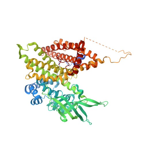
7S7G - PubMed Abstract:
Very long-chain acyl-CoA dehydrogenase (VLCAD) is an inner mitochondrial membrane enzyme that catalyzes the first and rate-limiting step of long-chain fatty acid oxidation. Point mutations in human VLCAD can produce an inborn error of metabolism called VLCAD deficiency that can lead to severe pathophysiologic consequences, including cardiomyopathy, hypoglycemia, and rhabdomyolysis. Discrete mutations in a structurally-uncharacterized C-terminal domain region of VLCAD cause enzymatic deficiency by an incompletely defined mechanism. Here, we conducted a structure-function study, incorporating X-ray crystallography, hydrogen-deuterium exchange mass spectrometry, computational modeling, and biochemical analyses, to characterize a specific membrane interaction defect of full-length, human VLCAD bearing the clinically-observed mutations, A450P or L462P. By disrupting a predicted α-helical hairpin, these mutations either partially or completely impair direct interaction with the membrane itself. Thus, our data support a structural basis for VLCAD deficiency in patients with discrete mutations in an α-helical membrane-binding motif, resulting in pathologic enzyme mislocalization.
- Department of Pediatric Oncology, Dana-Farber Cancer Institute, Boston, MA, USA.
Organizational Affiliation: