


Crystal structure of UrtA from Synechococcus CC9311 in complex with urea and calcium
Shah, B.S., Mikolajek, H., Mykhaylyk, V., Orr, C.M., Owens, R., Paulsen, I.T.To be published.
Experimental Data Snapshot
Entity ID: 1 | |||||
|---|---|---|---|---|---|
| Molecule | Chains | Sequence Length | Organism | Details | Image |
| Urea ABC transporter, periplasmic urea-binding protein | 400 | Synechococcus sp. CC9311 | Mutation(s): 0 Gene Names: sync_2872 | 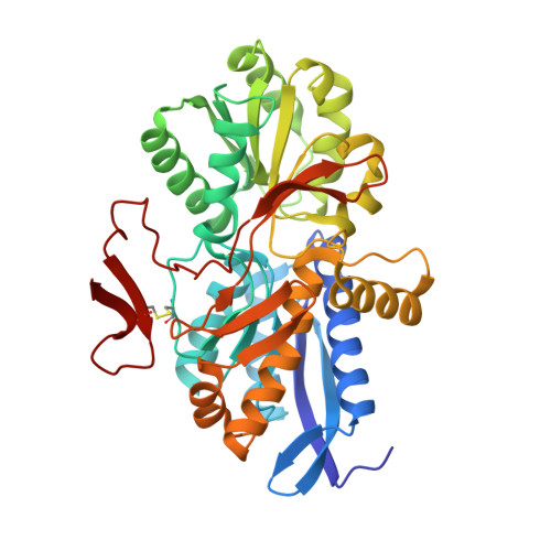 | |
| Ligands 4 Unique | |||||
|---|---|---|---|---|---|
| ID | Chains | Name / Formula / InChI Key | 2D Diagram | 3D Interactions | |
| EDO Download:Ideal Coordinates CCD File | D [auth A] E [auth A] F [auth A] G [auth A] H [auth A] | 1,2-ETHANEDIOL C2 H6 O2 LYCAIKOWRPUZTN-UHFFFAOYSA-N |  | ||
| URE (Subject of Investigation/LOI) Download:Ideal Coordinates CCD File | C [auth A], R [auth B] | UREA C H4 N2 O XSQUKJJJFZCRTK-UHFFFAOYSA-N |  | ||
| CA (Subject of Investigation/LOI) Download:Ideal Coordinates CCD File | K [auth A] L [auth A] M [auth A] N [auth A] O [auth A] | CALCIUM ION Ca BHPQYMZQTOCNFJ-UHFFFAOYSA-N |  | ||
| CL Download:Ideal Coordinates CCD File | AA [auth B] | CHLORIDE ION Cl VEXZGXHMUGYJMC-UHFFFAOYSA-M |  | ||
| Length ( Å ) | Angle ( ˚ ) |
|---|---|
| a = 49.76 | α = 90 |
| b = 117.12 | β = 90 |
| c = 123.46 | γ = 90 |
| Software Name | Purpose |
|---|---|
| REFMAC | refinement |
| XDS | data reduction |
| Coot | model building |
| CRANK2 | phasing |
| XSCALE | data scaling |
| Funding Organization | Location | Grant Number |
|---|---|---|
| Australian Research Council (ARC) | Australia | DP200102944 |
| Australian Research Council (ARC) | Australia | CE200100029 |


