Structural Characterization of Cytochrome c ' beta-Met from an Ammonia-Oxidizing Bacterium.
Abendroth, J., Buchko, G.W., Liew, F.N., Nguyen, J.N., Kim, H.J.(2022) Biochemistry 61: 563-574
- PubMed: 35315646 Search on PubMed
- DOI: https://doi.org/10.1021/acs.biochem.1c00640
- Primary Citation Related Structures:
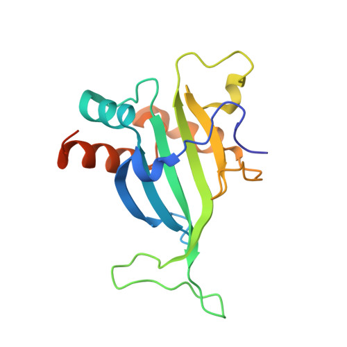
7S5O - PubMed Abstract:
The ammonia-oxidizing bacterium Nitrosomonas europaea expresses two cytochromes in the P460 superfamily that are predicted to be structurally similar. In one, cytochrome (cyt) P460, the substrate hydroxylamine (NH 2 OH) is converted to nitric oxide (NO) and nitrous oxide (N 2 O) requiring a unique heme-lysyl cross-link in the catalytic cofactor. In the second, cyt
' β-Met , the cross-link is absent, and the cytochrome instead binds H 2 O 2 forming a ferryl species similar to compound II of peroxidases. Here, we report the 1.80 Å crystal structure of cyt' β-Met ─a well-expressed protein in N. europaea with a lysine to a methionine replacement at the cross-linking position. The structure of cyt' β-Met is characterized by a large β-sheet typical of P460 members; however, several localized structural differences render cyt' β-Met distinct. This includes a large lasso-like loop at the "top" of the cytochrome that is not observed in other structurally characterized members. Active site variation is also observed, especially in comparison to its closest homologue cyt' β from the methane-oxidizing Methylococcus capsulatus Bath, which also lacks the cross-link. The phenylalanine "cap" which is presumed to control small ligand access to the distal heme iron is replaced with an arginine, reminiscent of the strictly conserved distal arginine in peroxidases and to the NH 2 OH-oxidizing cytochromes P460. A critical proton-transferring glutamate residue required for NH 2 OH oxidation is nevertheless missing in the active site. This in part explains the inability of cyt' β-Met to oxidize NH 2 OH. Our structure also rationalizes the absence of a methionyl cross-link, although the side chain's spatial position in the structure does not eliminate the possibility that it could form under certain conditions. - Seattle Structural Genomics Center for Infectious Diseases, Seattle, Washington 98105, United States.
Organizational Affiliation: