7S3L

Structure of Bacillus subtilis CcrZ (previously called YtmP)

  • Classification: CELL CYCLE
  • Organism(s): Bacillus subtilis PY79
  • Expression System: Escherichia coli
  • Mutation(s): No 

  • Deposited: 2021-09-07 Released: 2022-05-11 
  • Deposition Author(s): Nandakumar, J., Wozniak, K.J.
  • Funding Organization(s): National Institutes of Health/National Institute of General Medical Sciences (NIH/NIGMS), National Science Foundation (NSF, United States)

Experimental Data Snapshot

  • Method: X-RAY DIFFRACTION
  • Resolution: 2.60 Å
  • R-Value Free: 0.263 
  • R-Value Work: 0.242 
  • R-Value Observed: 0.244 

wwPDB Validation   3D Report Full Report


Ligand Structure Quality Assessment 


This is version 1.1 of the entry. See complete history


Literature

Structure and kinase activity of bacterial cell cycle regulator CcrZ.

Wozniak, K.J.Burby, P.E.Nandakumar, J.Simmons, L.A.

(2022) PLoS Genet 18: e1010196-e1010196

  • DOI: https://doi.org/10.1371/journal.pgen.1010196
  • Primary Citation of Related Structures:  
    7S3L

  • PubMed Abstract: 

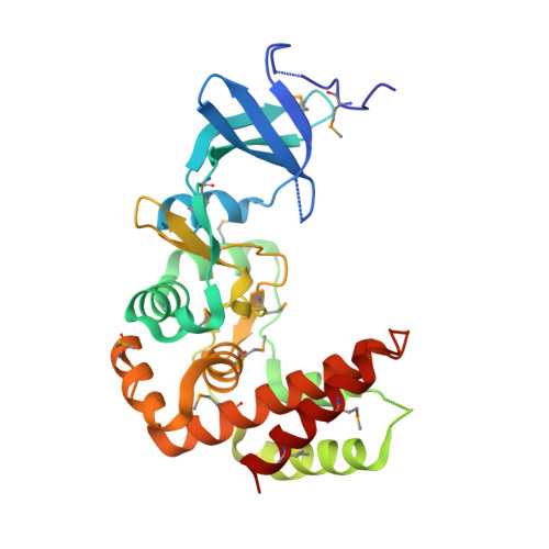
    CcrZ is a recently discovered cell cycle regulator that connects DNA replication initiation with cell division in pneumococci and may have a similar function in related bacteria. CcrZ is also annotated as a putative kinase, suggesting that CcrZ homologs could represent a novel family of bacterial kinase-dependent cell cycle regulators. Here, we investigate the CcrZ homolog in Bacillus subtilis and show that cells lacking ccrZ are sensitive to a broad range of DNA damage. We demonstrate that increased expression of ccrZ results in over-initiation of DNA replication. In addition, increased expression of CcrZ activates the DNA damage response. Using sensitivity to DNA damage as a proxy, we show that the negative regulator for replication initiation (yabA) and ccrZ function in the same pathway. We show that CcrZ interacts with replication initiation proteins DnaA and DnaB, further suggesting that CcrZ is important for replication timing. To understand how CcrZ functions, we solved the crystal structure bound to AMP-PNP to 2.6 Å resolution. The CcrZ structure most closely resembles choline kinases, consisting of a bilobal structure with a cleft between the two lobes for binding ATP and substrate. Inspection of the structure reveals a major restructuring of the substrate-binding site of CcrZ relative to the choline-binding pocket of choline kinases, consistent with our inability to detect activity with choline for this protein. Instead, CcrZ shows activity on D-ribose and 2-deoxy-D-ribose, indicating adaptation of the choline kinase fold in CcrZ to phosphorylate a novel substrate. We show that integrity of the kinase active site is required for ATPase activity in vitro and for function in vivo. This work provides structural, biochemical, and functional insight into a newly identified, and conserved group of bacterial kinases that regulate DNA replication initiation.


  • Organizational Affiliation

    Department of Molecular, Cellular, and Developmental Biology, University of Michigan, Ann Arbor, Michigan, United States of America.


Macromolecules
Find similar proteins by:  (by identity cutoff)  |  3D Structure
Entity ID: 1
MoleculeChains Sequence LengthOrganismDetailsImage
Aminoglycoside phosphotransferase family protein269Bacillus subtilis PY79Mutation(s): 0 
Gene Names: ytmP
UniProt
Find proteins for C0SPC1 (Bacillus subtilis (strain 168))
Explore C0SPC1 
Go to UniProtKB:  C0SPC1
Entity Groups  
Sequence Clusters30% Identity50% Identity70% Identity90% Identity95% Identity100% Identity
UniProt GroupC0SPC1
Sequence Annotations
Expand
  • Reference Sequence
Small Molecules
Ligands 1 Unique
IDChains Name / Formula / InChI Key2D Diagram3D Interactions
ANP (Subject of Investigation/LOI)
Query on ANP

Download Ideal Coordinates CCD File 
B [auth A]PHOSPHOAMINOPHOSPHONIC ACID-ADENYLATE ESTER
C10 H17 N6 O12 P3
PVKSNHVPLWYQGJ-KQYNXXCUSA-N
Modified Residues  1 Unique
IDChains TypeFormula2D DiagramParent
MSE
Query on MSE
A
L-PEPTIDE LINKINGC5 H11 N O2 SeMET
Experimental Data & Validation

Experimental Data

  • Method: X-RAY DIFFRACTION
  • Resolution: 2.60 Å
  • R-Value Free: 0.263 
  • R-Value Work: 0.242 
  • R-Value Observed: 0.244 
  • Space Group: P 43 21 2
Unit Cell:
Length ( Å )Angle ( ˚ )
a = 98.118α = 90
b = 98.118β = 90
c = 69.127γ = 90
Software Package:
Software NamePurpose
PHENIXrefinement
Cootmodel building
DIALSdata reduction
Aimlessdata scaling
AutoSolphasing

Structure Validation

View Full Validation Report


Ligand Structure Quality Assessment 


Entry History & Funding Information

Deposition Data


Funding OrganizationLocationGrant Number
National Institutes of Health/National Institute of General Medical Sciences (NIH/NIGMS)United StatesR35GM131772
National Institutes of Health/National Institute of General Medical Sciences (NIH/NIGMS)United StatesR01GM120094
National Institutes of Health/National Institute of General Medical Sciences (NIH/NIGMS)United StatesT32GM008353
National Science Foundation (NSF, United States)United StatesDEG1256260)

Revision History  (Full details and data files)

  • Version 1.0: 2022-05-11
    Type: Initial release
  • Version 1.1: 2022-06-01
    Changes: Database references