7RRK | pdb_00007rrk

Crystal structure of fast switching M159E mutant of fluorescent protein Dronpa (Dronpa2)


Experimental Data Snapshot

  • Method: X-RAY DIFFRACTION
  • Resolution: 1.93 Å
  • R-Value Free: 
    0.266 (Depositor), 0.267 (DCC) 
  • R-Value Work: 
    0.242 (Depositor), 0.244 (DCC) 
  • R-Value Observed: 
    0.243 (Depositor) 

Starting Model: experimental
View more details

wwPDB Validation 3D Report Full Report

Validation slider image for 7RRK

This is version 3.0 of the entry. See complete history

Literature

Energetic Basis and Design of Enzyme Function Demonstrated Using GFP, an Excited-State Enzyme.

Lin, C.Y.Romei, M.G.Mathews, I.I.Boxer, S.G.

(2022) J Am Chem Soc 144: 3968-3978

  • DOI: https://doi.org/10.1021/jacs.1c12305
  • Primary Citation Related Structures: 
    7RRH, 7RRI, 7RRJ, 7RRK

  • PubMed Abstract: 

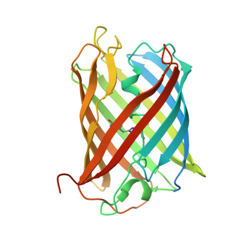
    The past decades have witnessed an explosion of de novo protein designs with a remarkable range of scaffolds. It remains challenging, however, to design catalytic functions that are competitive with naturally occurring counterparts as well as biomimetic or nonbiological catalysts. Although directed evolution often offers efficient solutions, the fitness landscape remains opaque. Green fluorescent protein (GFP), which has revolutionized biological imaging and assays, is one of the most redesigned proteins. While not an enzyme in the conventional sense, GFPs feature competing excited-state decay pathways with the same steric and electrostatic origins as conventional ground-state catalysts, and they exert exquisite control over multiple reaction outcomes through the same principles. Thus, GFP is an "excited-state enzyme". Herein we show that rationally designed mutants and hybrids that contain environmental mutations and substituted chromophores provide the basis for a quantitative model and prediction that describes the influence of sterics and electrostatics on excited-state catalysis of GFPs. As both perturbations can selectively bias photoisomerization pathways, GFPs with fluorescence quantum yields (FQYs) and photoswitching characteristics tailored for specific applications could be predicted and then demonstrated. The underlying energetic landscape, readily accessible via spectroscopy for GFPs, offers an important missing link in the design of protein function that is generalizable to catalyst design.


  • Organizational Affiliation
    • Department of Chemistry, Stanford University, Stanford, California 94305, United States.

Macromolecule Content 

  • Total Structure Weight: 231.92 kDa 
  • Atom Count: 15,363 
  • Modeled Residue Count: 1,727 
  • Deposited Residue Count: 2,040 
  • Unique protein chains: 1

Macromolecules

Find similar proteins by:|  3D Structure
Entity ID: 1
MoleculeChains  Sequence LengthOrganismDetailsImage
Fluorescent protein Dronpa
A, B, C, D, E
A, B, C, D, E, F, G, H
255Echinophyllia sp. SC22Mutation(s): 2 
Gene Names: Dronpa
UniProt
Find proteins for Q5TLG6 (Echinophyllia sp. SC22)
Explore Q5TLG6 
Go to UniProtKB:  Q5TLG6
Entity Groups
Sequence Clusters30% Identity50% Identity70% Identity90% Identity95% Identity100% Identity
UniProt GroupQ5TLG6
Sequence Annotations
Expand
Reference Sequence

Small Molecules

Modified Residues  1 Unique
IDChains TypeFormula2D DiagramParent
GYC
Query on GYC
A, B, C, D, E
A, B, C, D, E, F, G, H
L-PEPTIDE LINKINGC14 H15 N3 O4 SCYS, TYR, GLY

Experimental Data & Validation

Experimental Data

  • Method: X-RAY DIFFRACTION
  • Resolution: 1.93 Å
  • R-Value Free:  0.266 (Depositor), 0.267 (DCC) 
  • R-Value Work:  0.242 (Depositor), 0.244 (DCC) 
  • R-Value Observed: 0.243 (Depositor) 
Space Group: P 1 21 1
Unit Cell:
Length ( Å )Angle ( ˚ )
a = 79.825α = 90
b = 85.732β = 95.05
c = 143.05γ = 90
Software Package:
Software NamePurpose
PHENIXrefinement
XDSdata reduction
XDSdata scaling
PHASERphasing

Structure Validation

View Full Validation Report


Entry History 

& Funding Information

Deposition Data


Funding OrganizationLocationGrant Number
National Institutes of Health/National Institute of General Medical Sciences (NIH/NIGMS)United StatesGM118044
National Science Foundation (NSF, United States)United StatesCHE-1740645

Revision History  (Full details and data files)

  • Version 1.0: 2021-10-13
    Type: Initial release
  • Version 1.1: 2022-03-23
    Changes: Database references
  • Version 1.2: 2023-10-18
    Changes: Data collection, Refinement description
  • Version 2.0: 2023-11-15
    Changes: Atomic model, Data collection, Derived calculations
  • Version 2.1: 2024-11-13
    Changes: Structure summary
  • Version 3.0: 2026-03-18
    Changes: Polymer sequence