7RKB | pdb_00007rkb

Crystal Structure of Putative Pterin Binding Protein (PruR) from Klebsiella pneumoniae in Complex with Neopterin


Experimental Data Snapshot

  • Method: X-RAY DIFFRACTION
  • Resolution: 2.50 Å
  • R-Value Free: 
    0.233 (Depositor), 0.249 (DCC) 
  • R-Value Work: 
    0.188 (Depositor), 0.202 (DCC) 
  • R-Value Observed: 
    0.193 (Depositor) 

wwPDB Validation   3D Report Full Report


Ligand Structure Quality Assessment 


This is version 1.2 of the entry. See complete history


Literature

Control of biofilm formation by an Agrobacterium tumefaciens pterin-binding periplasmic protein conserved among diverse Proteobacteria.

Greenwich, J.L.Eagan, J.L.Feirer, N.Boswinkle, K.Minasov, G.Shuvalova, L.Inniss, N.L.Raghavaiah, J.Ghosh, A.K.Satchell, K.J.F.Allen, K.D.Fuqua, C.

(2024) Proc Natl Acad Sci U S A 121: e2319903121-e2319903121

  • DOI: https://doi.org/10.1073/pnas.2319903121
  • Primary Citation Related Structures: 
    7KOM, 7KOS, 7KOU, 7KP2, 7RKB

  • PubMed Abstract: 

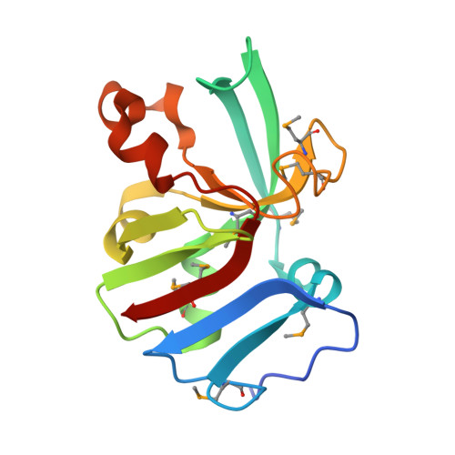
    Biofilm formation and surface attachment in multiple Alphaproteobacteria is driven by unipolar polysaccharide (UPP) adhesins. The pathogen Agrobacterium tumefaciens produces a UPP adhesin, which is regulated by the intracellular second messenger cyclic diguanylate monophosphate (c-di-GMP). Prior studies revealed that DcpA, a diguanylate cyclase-phosphodiesterase, is crucial in control of UPP production and surface attachment. DcpA is regulated by PruR, a protein with distant similarity to enzymatic domains known to coordinate the molybdopterin cofactor (MoCo). Pterins are bicyclic nitrogen-rich compounds, several of which are produced via a nonessential branch of the folate biosynthesis pathway, distinct from MoCo. The pterin-binding protein PruR controls DcpA activity, fostering c-di-GMP breakdown and dampening its synthesis. Pterins are excreted, and we report here that PruR associates with these metabolites in the periplasm, promoting interaction with the DcpA periplasmic domain. The pteridine reductase PruA, which reduces specific dihydro-pterin molecules to their tetrahydro forms, imparts control over DcpA activity through PruR. Tetrahydromonapterin preferentially associates with PruR relative to other related pterins, and the PruR-DcpA interaction is decreased in a pruA mutant. PruR and DcpA are encoded in an operon with wide conservation among diverse Proteobacteria including mammalian pathogens. Crystal structures reveal that PruR and several orthologs adopt a conserved fold, with a pterin-specific binding cleft that coordinates the bicyclic pterin ring. These findings define a pterin-responsive regulatory mechanism that controls biofilm formation and related c-di-GMP-dependent phenotypes in A. tumefaciens and potentially acts more widely in multiple proteobacterial lineages.


  • Organizational Affiliation
    • Department of Biology, Indiana University, Bloomington, IN 47405.

Macromolecules
Find similar proteins by:  (by identity cutoff)  |  3D Structure
Entity ID: 1
MoleculeChains Sequence LengthOrganismDetailsImage
Pterin Binding Protein149Klebsiella pneumoniae subsp. pneumoniae SA1Mutation(s): 0 
Gene Names: RJA_09115
Entity Groups  
Sequence Clusters30% Identity50% Identity70% Identity90% Identity95% Identity100% Identity
Sequence Annotations
Expand
  • Reference Sequence
Small Molecules
Ligands 3 Unique
IDChains Name / Formula / InChI Key2D Diagram3D Interactions
NEU (Subject of Investigation/LOI)
Query on NEU

Download Ideal Coordinates CCD File 
B [auth A]L-NEOPTERIN
C9 H11 N5 O4
BMQYVXCPAOLZOK-XINAWCOVSA-N
SO4
Query on SO4

Download Ideal Coordinates CCD File 
H [auth A],
I [auth A],
J [auth A],
K [auth A]
SULFATE ION
O4 S
QAOWNCQODCNURD-UHFFFAOYSA-L
CL
Query on CL

Download Ideal Coordinates CCD File 
C [auth A],
D [auth A],
E [auth A],
F [auth A],
G [auth A]
CHLORIDE ION
Cl
VEXZGXHMUGYJMC-UHFFFAOYSA-M
Modified Residues  1 Unique
IDChains TypeFormula2D DiagramParent
MSE
Query on MSE
A
L-PEPTIDE LINKINGC5 H11 N O2 SeMET
Experimental Data & Validation

Experimental Data

  • Method: X-RAY DIFFRACTION
  • Resolution: 2.50 Å
  • R-Value Free:  0.233 (Depositor), 0.249 (DCC) 
  • R-Value Work:  0.188 (Depositor), 0.202 (DCC) 
  • R-Value Observed: 0.193 (Depositor) 
Space Group: P 41 21 2
Unit Cell:
Length ( Å )Angle ( ˚ )
a = 83.521α = 90
b = 83.521β = 90
c = 45.884γ = 90
Software Package:
Software NamePurpose
REFMACrefinement
PDB_EXTRACTdata extraction
HKL-3000data reduction
HKL-3000data scaling
HKL-3000phasing

Structure Validation

View Full Validation Report


Ligand Structure Quality Assessment 


Entry History & Funding Information

Deposition Data


Funding OrganizationLocationGrant Number
National Institutes of Health/National Institute Of Allergy and Infectious Diseases (NIH/NIAID)United States--

Revision History  (Full details and data files)

  • Version 1.0: 2022-08-10
    Type: Initial release
  • Version 1.1: 2024-11-13
    Changes: Data collection, Structure summary
  • Version 1.2: 2026-04-01
    Changes: Database references