


Crystal structure of the Vitronectin hemopexin-like domain binding Calcium
Aleshin, A.E., Marassi, F.M.To be published.
Experimental Data Snapshot
Starting Model: experimental
View more details
Entity ID: 1 | |||||
|---|---|---|---|---|---|
| Molecule | Chains | Sequence Length | Organism | Details | Image |
| Vitronectin | 204 | Homo sapiens | Mutation(s): 2 Gene Names: VTN | 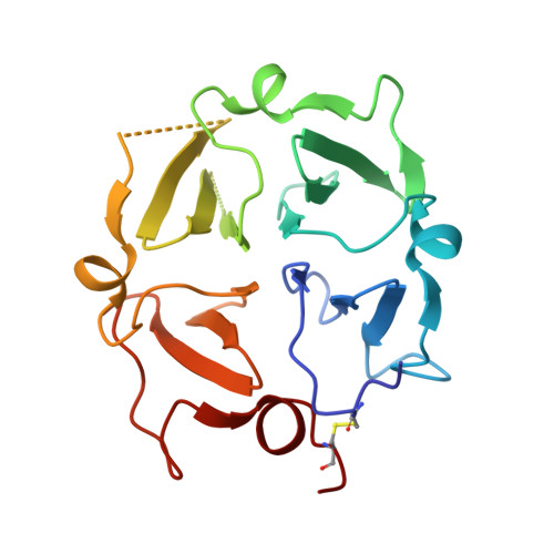 | |
UniProt & NIH Common Fund Data Resources | |||||
PHAROS: P04004 GTEx: ENSG00000109072 | |||||
Entity Groups | |||||
| Sequence Clusters | 30% Identity50% Identity70% Identity90% Identity95% Identity100% Identity | ||||
| UniProt Group | P04004 | ||||
Sequence AnnotationsExpand | |||||
Reference Sequence | |||||
| Ligands 4 Unique | |||||
|---|---|---|---|---|---|
| ID | Chains | Name / Formula / InChI Key | 2D Diagram | 3D Interactions | |
| SO4 (Subject of Investigation/LOI) Download:Ideal Coordinates CCD File | I [auth B] | SULFATE ION O4 S QAOWNCQODCNURD-UHFFFAOYSA-L |  | ||
| CA (Subject of Investigation/LOI) Download:Ideal Coordinates CCD File | C [auth A], F [auth B] | CALCIUM ION Ca BHPQYMZQTOCNFJ-UHFFFAOYSA-N |  | ||
| CL (Subject of Investigation/LOI) Download:Ideal Coordinates CCD File | D [auth A], G [auth B] | CHLORIDE ION Cl VEXZGXHMUGYJMC-UHFFFAOYSA-M |  | ||
| NA (Subject of Investigation/LOI) Download:Ideal Coordinates CCD File | E [auth A], H [auth B] | SODIUM ION Na FKNQFGJONOIPTF-UHFFFAOYSA-N |  | ||
| Length ( Å ) | Angle ( ˚ ) |
|---|---|
| a = 40.832 | α = 90 |
| b = 125.575 | β = 119.25 |
| c = 40.883 | γ = 90 |
| Software Name | Purpose |
|---|---|
| REFMAC | refinement |
| PDB_EXTRACT | data extraction |
| XDS | data reduction |
| Aimless | data scaling |
| PHASER | phasing |
| Funding Organization | Location | Grant Number |
|---|---|---|
| National Institutes of Health/Eunice Kennedy Shriver National Institute of Child Health & Human Development (NIH/NICHD) | United States | -- |


