7R0X | pdb_00007r0x

Structure of the branching thioesterase from oocydin biosynthesis


Experimental Data Snapshot

  • Method: X-RAY DIFFRACTION
  • Resolution: 2.83 Å
  • R-Value Free: 
    0.281 (Depositor), 0.274 (DCC) 
  • R-Value Work: 
    0.224 (Depositor), 0.224 (DCC) 
  • R-Value Observed: 
    0.229 (Depositor) 

wwPDB Validation 3D Report Full Report

Validation slider image for 7R0X

This is version 1.3 of the entry. See complete history

Literature

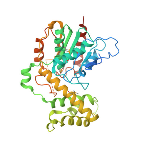
Structure of a Promiscuous Thioesterase Domain Responsible for Branching Acylation in Polyketide Biosynthesis.

Fraley, A.E.Dieterich, C.L.Mabesoone, M.F.J.Minas, H.A.Meoded, R.A.Hemmerling, F.Piel, J.

(2022) Angew Chem Int Ed Engl 61: e202206385-e202206385

  • DOI: https://doi.org/10.1002/anie.202206385
  • Primary Citation Related Structures: 
    7R0X

  • PubMed Abstract: 

    Thioesterases (TEs) are fundamentally important enzymes present in all bacteria and eukaryotes, where they have conserved functions in fatty acid biosynthesis and secondary metabolism. This work provides the first structural insights into a functionally distinct group of TEs that perform diverse acylations in polyketide and peptide biosynthesis (TE B s). Structural analysis of the oocydin (OocS) TE B domain facilitated identification and engineering of the active site to modulate acyl-group acceptance. In this way, we achieved higher reactivity using a structure-based approach, building a foundation for biocatalytic development of TE B -mediated O-acylation, a modification known to improve the bioactivity of oocydin-type polyketides. Lastly, the promiscuity of the OocS TE B motivated us to investigate, and ultimately provide evidence for, the production of longer chain branched oocydins in the native host Serratia plymuthica 4Rx13. This work frames the OocS TE B and homologs as invaluable synthetic biology tools for polyketide drug development.


  • Organizational Affiliation
    • Department of Biology, Institute of Microbiology, Eidgenössische Technische Hochschule (ETH) Zurich, Vladimir-Prelog-Weg 4, 8093, Zurich, Switzerland.

Macromolecule Content 

  • Total Structure Weight: 41.1 kDa 
  • Atom Count: 2,457 
  • Modeled Residue Count: 300 
  • Deposited Residue Count: 364 
  • Unique protein chains: 1

Macromolecules

Find similar proteins by:|  3D Structure
Entity ID: 1
MoleculeChains  Sequence LengthOrganismDetailsImage
Polyketide synthase364SerratiaMutation(s): 0 
Gene Names: CT690_01900
UniProt
Find proteins for A0A318P693 (Serratia plymuthica)
Explore A0A318P693 
Go to UniProtKB:  A0A318P693
Entity Groups
Sequence Clusters30% Identity50% Identity70% Identity90% Identity95% Identity100% Identity
UniProt GroupA0A318P693
Sequence Annotations
Expand
Reference Sequence

Small Molecules

Modified Residues  1 Unique
IDChains TypeFormula2D DiagramParent
MSE
Query on MSE
A
L-PEPTIDE LINKINGC5 H11 N O2 SeMET

Experimental Data & Validation

Experimental Data

  • Method: X-RAY DIFFRACTION
  • Resolution: 2.83 Å
  • R-Value Free:  0.281 (Depositor), 0.274 (DCC) 
  • R-Value Work:  0.224 (Depositor), 0.224 (DCC) 
  • R-Value Observed: 0.229 (Depositor) 
Space Group: I 4
Unit Cell:
Length ( Å )Angle ( ˚ )
a = 106.818α = 90
b = 106.818β = 90
c = 78.14γ = 90
Software Package:
Software NamePurpose
PHENIXrefinement
XDSdata scaling
Aimlessdata scaling
AutoSolphasing

Structure Validation

View Full Validation Report


Entry History 

& Funding Information

Deposition Data


Funding OrganizationLocationGrant Number
European Research Council (ERC)European Union742739

Revision History  (Full details and data files)

  • Version 1.0: 2022-08-10
    Type: Initial release
  • Version 1.1: 2022-08-31
    Changes: Database references
  • Version 1.2: 2022-09-28
    Changes: Database references
  • Version 1.3: 2024-11-06
    Changes: Data collection, Structure summary