Serial Femtosecond Crystallography Reveals the Role of Water in the One- or Two-Electron Redox Chemistry of Compound I in the Catalytic Cycle of the B-Type Dye-Decolorizing Peroxidase DtpB.
Lucic, M., Wilson, M.T., Tosha, T., Sugimoto, H., Shilova, A., Axford, D., Owen, R.L., Hough, M.A., Worrall, J.A.R.(2022) ACS Catal 12: 13349-13359
- PubMed: 36366763 Search on PubMedSearch on PubMed Central
- DOI: https://doi.org/10.1021/acscatal.2c03754
- Primary Citation Related Structures:
7QZE, 7QZF, 7QZG, 7QZH, 7ZMJ - PubMed Abstract:
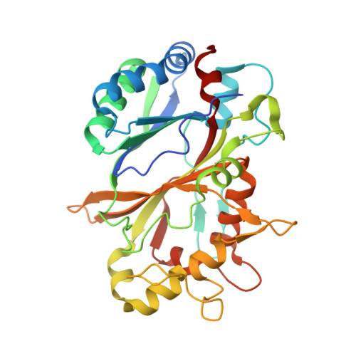
Controlling the reactivity of high-valent Fe(IV)-O catalytic intermediates, Compounds I and II, generated in heme enzymes upon reaction with dioxygen or hydrogen peroxide, is important for function. It has been hypothesized that the presence (wet) or absence (dry) of distal heme pocket water molecules can influence whether Compound I undergoes sequential one-electron additions or a concerted two-electron reduction. To test this hypothesis, we investigate the role of water in the heme distal pocket of a dye-decolorizing peroxidase utilizing a combination of serial femtosecond crystallography and rapid kinetic studies. In a dry distal heme site, Compound I reduction proceeds through a mechanism in which Compound II concentration is low. This reaction shows a strong deuterium isotope effect, indicating that reduction is coupled to proton uptake. The resulting protonated Compound II (Fe(IV)-OH) rapidly reduces to the ferric state, giving the appearance of a two-electron transfer process. In a wet site, reduction of Compound I is faster, has no deuterium effect, and yields highly populated Compound II, which is subsequently reduced to the ferric form. This work provides a definitive experimental test of the hypothesis advanced in the literature that relates sequential or concerted electron transfer to Compound I in wet or dry distal heme sites.
- School of Life Sciences, University of Essex, Wivenhoe Park, Essex, ColchesterCO4 3SQ, U.K.
Organizational Affiliation: