Structural Characterization of Lytic Transglycosylase SltB2 of Pseudomonas aeruginosa.
Miguel-Ruano, V., Batuecas, M.T., Lastochkin, E., Dominguez-Gil, T., Molina, R., Mobashery, S., Hermoso, J.A.(2025) ACS Omega 10: 48385-48394
- PubMed: 41141816 Search on PubMedSearch on PubMed Central
- DOI: https://doi.org/10.1021/acsomega.5c05747
- Primary Citation Related Structures:
7QVD - PubMed Abstract:
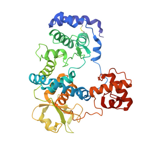
Lytic transglycosylases (LTs) belong to a family of enzymes that turnover the bacterial cell-wall peptidoglycan through a nonhydrolytic cleavage of the β(1-4) glycosidic bond, generating a hallmark 1,6-anhydromuramyl moiety in the reaction products. LTs are essential for numerous cellular processes, including cell-wall maturation, peptidoglycan recycling, cell division, and the assembly of multiprotein complexes. Their functional diversity underscores their biological significance. Family 3 LTs are distinguished by their EF-hand Ca 2+ -binding motif and are classified into two subfamilies. Subfamily 3B members, including Pseudomonas aeruginosa SltB2, possess a peptidoglycan-binding domain absent in subfamily 3A. In this study, we present the structural characterization of P. aeruginosa SltB2. The high-resolution crystal structure of SltB2 reveals a unique modular architecture shaped by the specific arrangement of its PG-binding domain and distinct differences in the organization of key residues surrounding the catalytic Glu residue compared to other family 3 members. A model of interaction between SltB2 and the peptidoglycan is proposed, which accounts for the enzyme's tolerance to peptide stems and reveals particular features at site +2, due to the unique arrangement of the PG-binding domain, explaining its preferred exolytic activity. Comparative structural analyses of Family 3 LTs provide insights into substrate recognition and enzymatic function, advancing our understanding of bacterial cell-wall remodeling mechanisms.
- Department of Crystallography and Structural Biology, Institute of Physical-Chemistry "Blas Cabrera", Spanish National Research Council (CSIC), Serrano 119, Madrid 28006, Spain.
Organizational Affiliation: