7QSR

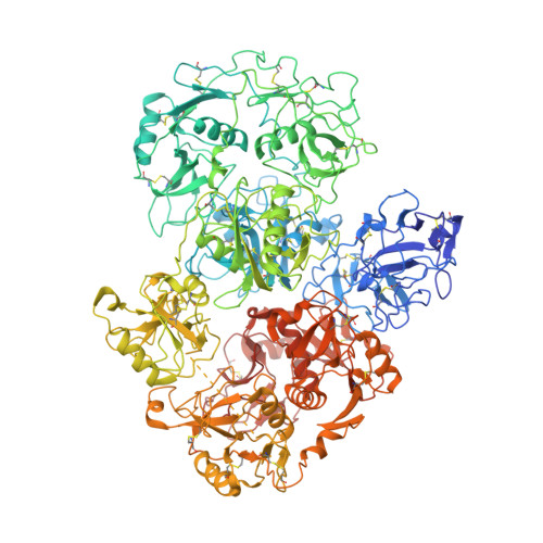
CryoEM structure of the Ectodomain of Human PLA2R


Experimental Data Snapshot

  • Method: ELECTRON MICROSCOPY
  • Resolution: 3.40 Å
  • Aggregation State: 2D ARRAY 
  • Reconstruction Method: SINGLE PARTICLE 

Starting Model: experimental
View more details

wwPDB Validation   3D Report Full Report


This is version 1.2 of the entry. See complete history


Literature

Structure of PLA2R reveals presentation of the dominant membranous nephropathy epitope and an immunogenic patch.

Fresquet, M.Lockhart-Cairns, M.P.Rhoden, S.J.Jowitt, T.A.Briggs, D.C.Baldock, C.Brenchley, P.E.Lennon, R.

(2022) Proc Natl Acad Sci U S A 119: e2202209119-e2202209119

  • DOI: https://doi.org/10.1073/pnas.2202209119
  • Primary Citation of Related Structures:  
    7QSR

  • PubMed Abstract: 

    Membranous nephropathy is an autoimmune kidney disease caused by autoantibodies targeting antigens present on glomerular podocytes, instigating a cascade leading to glomerular injury. The most prevalent circulating autoantibodies in membranous nephropathy are against phospholipase A2 receptor (PLA2R), a cell surface receptor. The dominant epitope in PLA2R is located within the cysteine-rich domain, yet high-resolution structure-based mapping is lacking. In this study, we define the key nonredundant amino acids in the dominant epitope of PLA2R involved in autoantibody binding. We further describe two essential regions within the dominant epitope and spacer requirements for a synthetic peptide of the epitope for drug discovery. In addition, using cryo-electron microscopy, we have determined the high-resolution structure of PLA2R to 3.4 Å resolution, which shows that the dominant epitope and key residues within the cysteine-rich domain are accessible at the cell surface. In addition, the structure of PLA2R not only suggests a different orientation of domains but also implicates a unique immunogenic signature in PLA2R responsible for inducing autoantibody formation and recognition.


  • Organizational Affiliation

    Wellcome Centre for Cell-Matrix Research, The University of Manchester, Manchester, M13 9PT, United Kingdom.


Macromolecules
Find similar proteins by:  (by identity cutoff)  |  3D Structure
Entity ID: 1
MoleculeChains Sequence LengthOrganismDetailsImage
Secretory phospholipase A2 receptor1,377Homo sapiensMutation(s): 0 
Gene Names: PLA2R1CLEC13C
UniProt & NIH Common Fund Data Resources
Find proteins for Q13018 (Homo sapiens)
Explore Q13018 
Go to UniProtKB:  Q13018
PHAROS:  Q13018
GTEx:  ENSG00000153246 
Entity Groups  
Sequence Clusters30% Identity50% Identity70% Identity90% Identity95% Identity100% Identity
UniProt GroupQ13018
Glycosylation
Glycosylation Sites: 13Go to GlyGen: Q13018-1
Sequence Annotations
Expand
  • Reference Sequence
Oligosaccharides

Help

Entity ID: 2
MoleculeChains Length2D Diagram Glycosylation3D Interactions
beta-D-mannopyranose-(1-4)-2-acetamido-2-deoxy-beta-D-glucopyranose-(1-4)-2-acetamido-2-deoxy-beta-D-glucopyranose
B, C
3N-Glycosylation
Glycosylation Resources
GlyTouCan:  G15407YE
GlyCosmos:  G15407YE
GlyGen:  G15407YE
Entity ID: 3
MoleculeChains Length2D Diagram Glycosylation3D Interactions
alpha-D-mannopyranose-(1-3)-beta-D-mannopyranose-(1-4)-2-acetamido-2-deoxy-beta-D-glucopyranose-(1-4)-2-acetamido-2-deoxy-beta-D-glucopyranose
D
4N-Glycosylation
Glycosylation Resources
GlyTouCan:  G81315DD
GlyCosmos:  G81315DD
GlyGen:  G81315DD
Entity ID: 4
MoleculeChains Length2D Diagram Glycosylation3D Interactions
alpha-D-mannopyranose-(1-6)-beta-D-mannopyranose-(1-3)-2-acetamido-2-deoxy-beta-D-glucopyranose-(1-6)-2-acetamido-2-deoxy-beta-D-glucopyranose
E
4N-Glycosylation
Small Molecules
Ligands 1 Unique
IDChains Name / Formula / InChI Key2D Diagram3D Interactions
NAG
Query on NAG

Download Ideal Coordinates CCD File 
F [auth A]
G [auth A]
H [auth A]
I [auth A]
J [auth A]
F [auth A],
G [auth A],
H [auth A],
I [auth A],
J [auth A],
K [auth A],
L [auth A],
M [auth A],
N [auth A]
2-acetamido-2-deoxy-beta-D-glucopyranose
C8 H15 N O6
OVRNDRQMDRJTHS-FMDGEEDCSA-N
Experimental Data & Validation

Experimental Data

  • Method: ELECTRON MICROSCOPY
  • Resolution: 3.40 Å
  • Aggregation State: 2D ARRAY 
  • Reconstruction Method: SINGLE PARTICLE 
EM Software:
TaskSoftware PackageVersion
MODEL REFINEMENTPHENIX1.19.2

Structure Validation

View Full Validation Report


Entry History & Funding Information

Deposition Data


Funding OrganizationLocationGrant Number
European Union (EU)United Kingdom305608
Wellcome TrustUnited Kingdom097820
Wellcome TrustUnited Kingdom203128/Z/16/Z
Wellcome TrustUnited Kingdom202860/Z/16/Z
Wellcome TrustUnited Kingdom108466/Z/15/Z

Revision History  (Full details and data files)

  • Version 1.0: 2022-07-20
    Type: Initial release
  • Version 1.1: 2022-08-03
    Changes: Database references
  • Version 1.2: 2024-11-06
    Changes: Data collection, Refinement description, Structure summary