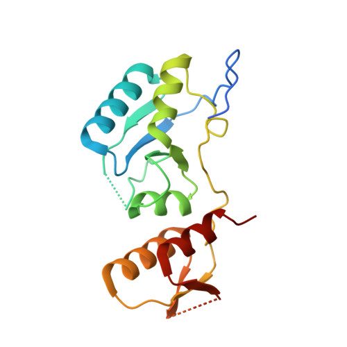
7QRO | pdb_00007qro

Crystal structure of the unconventional kinetochore protein Trypanosoma brucei KKT4 BRCT domain K543A mutant


Experimental Data Snapshot

  • Method: X-RAY DIFFRACTION
  • Resolution: 1.80 Å
  • R-Value Free: 
    0.231 (Depositor), 0.232 (DCC) 
  • R-Value Work: 
    0.196 (Depositor), 0.199 (DCC) 
  • R-Value Observed: 
    0.198 (Depositor) 

Starting Model: experimental
View more details

wwPDB Validation 3D Report Full Report

Validation slider image for 7QRO

This is version 1.2 of the entry. See complete history

Literature

NMR study of the structure and dynamics of the BRCT domain from the kinetochore protein KKT4.

Ludzia, P.Hayashi, H.Robinson, T.Akiyoshi, B.Redfield, C.

(2024) Biomol NMR Assign 18: 15-25

  • DOI: https://doi.org/10.1007/s12104-024-10163-9
  • Primary Citation Related Structures: 
    7QRO

  • PubMed Abstract: 

    KKT4 is a multi-domain kinetochore protein specific to kinetoplastids, such as Trypanosoma brucei. It lacks significant sequence similarity to known kinetochore proteins in other eukaryotes. Our recent X-ray structure of the C-terminal region of KKT4 shows that it has a tandem BRCT (BRCA1 C Terminus) domain fold with a sulfate ion bound in a typical binding site for a phosphorylated serine or threonine. Here we present the 1 H, 13 C and 15 N resonance assignments for the BRCT domain of KKT4 (KKT4 463-645 ) from T. brucei. We show that the BRCT domain can bind phosphate ions in solution using residues involved in sulfate ion binding in the X-ray structure. We have used these assignments to characterise the secondary structure and backbone dynamics of the BRCT domain in solution. Mutating the residues involved in phosphate ion binding in T. brucei KKT4 BRCT results in growth defects confirming the importance of the BRCT phosphopeptide-binding activity in vivo. These results may facilitate rational drug design efforts in the future to combat diseases caused by kinetoplastid parasites.


  • Organizational Affiliation
    • Department of Biochemistry, University of Oxford, South Parks Road, Oxford, OX1 3QU, UK.

Macromolecule Content 

  • Total Structure Weight: 19.85 kDa 
  • Atom Count: 1,388 
  • Modeled Residue Count: 157 
  • Deposited Residue Count: 183 
  • Unique protein chains: 1

Macromolecules

Find similar proteins by:|  3D Structure
Entity ID: 1
MoleculeChains  Sequence LengthOrganismDetailsImage
Trypanosoma brucei KKT4 463-645 K543A183Trypanosoma brucei bruceiMutation(s): 1 
Gene Names: Tb927.8.3680
UniProt
Find proteins for Q580Y8 (Trypanosoma brucei brucei (strain 927/4 GUTat10.1))
Explore Q580Y8 
Go to UniProtKB:  Q580Y8
Entity Groups
Sequence Clusters30% Identity50% Identity70% Identity90% Identity95% Identity100% Identity
UniProt GroupQ580Y8
Sequence Annotations
Expand
Reference Sequence

Experimental Data & Validation

Experimental Data

  • Method: X-RAY DIFFRACTION
  • Resolution: 1.80 Å
  • R-Value Free:  0.231 (Depositor), 0.232 (DCC) 
  • R-Value Work:  0.196 (Depositor), 0.199 (DCC) 
  • R-Value Observed: 0.198 (Depositor) 
Space Group: P 21 21 21
Unit Cell:
Length ( Å )Angle ( ˚ )
a = 46.663α = 90
b = 61.343β = 90
c = 67.745γ = 90
Software Package:
Software NamePurpose
PHENIXrefinement
DIALSdata reduction
DIALSdata scaling
PHASERphasing

Structure Validation

View Full Validation Report


Entry History 

& Funding Information

Deposition Data


Funding OrganizationLocationGrant Number
Wellcome TrustUnited Kingdom210622/Z/18/Z

Revision History  (Full details and data files)

  • Version 1.0: 2022-02-09
    Type: Initial release
  • Version 1.1: 2024-01-31
    Changes: Data collection, Refinement description
  • Version 1.2: 2024-05-22
    Changes: Database references