7QRF

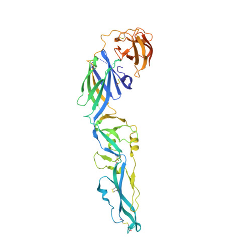
Structure of the dimeric complex between precursor membrane ectodomain (prM) and envelope protein ectodomain (E) from tick-borne encephalitis virus


Experimental Data Snapshot

  • Method: X-RAY DIFFRACTION
  • Resolution: 2.28 Å
  • R-Value Free: 0.213 
  • R-Value Work: 0.181 
  • R-Value Observed: 0.183 

Starting Models: experimental
View more details

wwPDB Validation   3D Report Full Report


This is version 1.3 of the entry. See complete history


Literature

Evolution and activation mechanism of the flavivirus class II membrane-fusion machinery.

Vaney, M.C.Dellarole, M.Duquerroy, S.Medits, I.Tsouchnikas, G.Rouvinski, A.England, P.Stiasny, K.Heinz, F.X.Rey, F.A.

(2022) Nat Commun 13: 3718-3718

  • DOI: https://doi.org/10.1038/s41467-022-31111-y
  • Primary Citation of Related Structures:  
    7QRE, 7QRF, 7QRG

  • PubMed Abstract: 

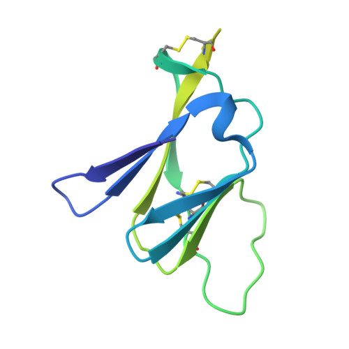
    The flavivirus envelope glycoproteins prM and E drive the assembly of icosahedral, spiky immature particles that bud across the membrane of the endoplasmic reticulum. Maturation into infectious virions in the trans-Golgi network involves an acid-pH-driven rearrangement into smooth particles made of (prM/E) 2 dimers exposing a furin site for prM cleavage into "pr" and "M". Here we show that the prM "pr" moiety derives from an HSP40 cellular chaperonin. Furthermore, the X-ray structure of the tick-borne encephalitis virus (pr/E) 2 dimer at acidic pH reveals the E 150-loop as a hinged-lid that opens at low pH to expose a positively-charged pr-binding pocket at the E dimer interface, inducing (prM/E) 2 dimer formation to generate smooth particles in the Golgi. Furin cleavage is followed by lid-closure upon deprotonation in the neutral-pH extracellular environment, expelling pr while the 150-loop takes the relay in fusion loop protection, thus revealing the elusive flavivirus mechanism of fusion activation.


  • Organizational Affiliation

    Institut Pasteur, Université Paris Cité, CNRS UMR 3569, Unité de Virologie Structurale, Paris, France.


Macromolecules
Find similar proteins by:  (by identity cutoff)  |  3D Structure
Entity ID: 1
MoleculeChains Sequence LengthOrganismDetailsImage
Envelope protein E443Tick-borne encephalitis virus (WESTERN SUBTYPE)Mutation(s): 0 
UniProt
Find proteins for P14336 (Tick-borne encephalitis virus European subtype (strain Neudoerfl))
Explore P14336 
Go to UniProtKB:  P14336
Entity Groups  
Sequence Clusters30% Identity50% Identity70% Identity90% Identity95% Identity100% Identity
UniProt GroupP14336
Sequence Annotations
Expand
  • Reference Sequence

Find similar proteins by:  Sequence   |   3D Structure  

Entity ID: 2
MoleculeChains Sequence LengthOrganismDetailsImage
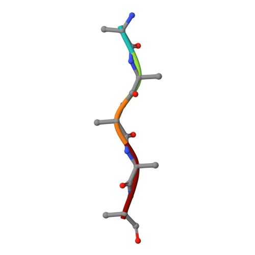
Unknown peptideB [auth C]5Tick-borne encephalitis virus (WESTERN SUBTYPE)Mutation(s): 0 
Sequence Annotations
Expand
  • Reference Sequence
Find similar proteins by:  (by identity cutoff)  |  3D Structure
Entity ID: 3
MoleculeChains Sequence LengthOrganismDetailsImage
Genome polyproteinC [auth D]138Tick-borne encephalitis virus (WESTERN SUBTYPE)Mutation(s): 0 
UniProt
Find proteins for P14336 (Tick-borne encephalitis virus European subtype (strain Neudoerfl))
Explore P14336 
Go to UniProtKB:  P14336
Entity Groups  
Sequence Clusters30% Identity50% Identity70% Identity90% Identity95% Identity100% Identity
UniProt GroupP14336
Sequence Annotations
Expand
  • Reference Sequence
Small Molecules
Ligands 3 Unique
IDChains Name / Formula / InChI Key2D Diagram3D Interactions
PE4
Query on PE4

Download Ideal Coordinates CCD File 
E [auth A]2-{2-[2-(2-{2-[2-(2-ETHOXY-ETHOXY)-ETHOXY]-ETHOXY}-ETHOXY)-ETHOXY]-ETHOXY}-ETHANOL
C16 H34 O8
PJWQOENWHPEPKI-UHFFFAOYSA-N
SO4
Query on SO4

Download Ideal Coordinates CCD File 
D [auth A]SULFATE ION
O4 S
QAOWNCQODCNURD-UHFFFAOYSA-L
EDO
Query on EDO

Download Ideal Coordinates CCD File 
F [auth A]
G [auth A]
H [auth A]
I [auth A]
J [auth A]
F [auth A],
G [auth A],
H [auth A],
I [auth A],
J [auth A],
K [auth A],
L [auth D],
M [auth D]
1,2-ETHANEDIOL
C2 H6 O2
LYCAIKOWRPUZTN-UHFFFAOYSA-N
Experimental Data & Validation

Experimental Data

  • Method: X-RAY DIFFRACTION
  • Resolution: 2.28 Å
  • R-Value Free: 0.213 
  • R-Value Work: 0.181 
  • R-Value Observed: 0.183 
  • Space Group: H 3 2
Unit Cell:
Length ( Å )Angle ( ˚ )
a = 155.73α = 90
b = 155.73β = 90
c = 161.88γ = 120
Software Package:
Software NamePurpose
BUSTERrefinement
PDB_EXTRACTdata extraction
XDSdata reduction
STARANISOdata scaling
PHASERphasing

Structure Validation

View Full Validation Report


Entry History & Funding Information

Deposition Data


Funding OrganizationLocationGrant Number
Pasteur InstituteFrance--
Centre National de la Recherche Scientifique (CNRS)France--
Agence Nationale de la Recherche (ANR)FranceANR-13-ISV8-0002-01

Revision History  (Full details and data files)

  • Version 1.0: 2022-05-25
    Type: Initial release
  • Version 1.1: 2022-12-07
    Changes: Database references
  • Version 1.2: 2024-01-31
    Changes: Data collection, Refinement description
  • Version 1.3: 2024-11-20
    Changes: Structure summary