


Discovery of Lu AF11167, a Phosphodiesterase 10A inhibitor clinical candidate
Kehler, J., Kilburn, J.P., Langgard, M., Christoffersen, C.T., Ritzen, A., Marigo, M., Jessing, M., Bundgaard, C., Puschl, A., Feigin, K., Nielsen, J.To be published.
Experimental Data Snapshot
Starting Model: experimental
View more details
Entity ID: 1 | |||||
|---|---|---|---|---|---|
| Molecule | Chains | Sequence Length | Organism | Details | Image |
| cAMP and cAMP-inhibited cGMP 3',5'-cyclic phosphodiesterase 10A | 340 | Homo sapiens | Mutation(s): 0 Gene Names: PDE10A EC: 3.1.4.17 | 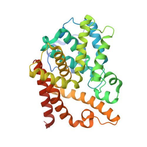 | |
UniProt & NIH Common Fund Data Resources | |||||
GTEx: ENSG00000112541 | |||||
Entity Groups | |||||
| Sequence Clusters | 30% Identity50% Identity70% Identity90% Identity95% Identity100% Identity | ||||
| UniProt Group | Q9Y233 | ||||
Sequence AnnotationsExpand | |||||
Reference Sequence | |||||
| Ligands 3 Unique | |||||
|---|---|---|---|---|---|
| ID | Chains | Name / Formula / InChI Key | 2D Diagram | 3D Interactions | |
| EHI (Subject of Investigation/LOI) Download:Ideal Coordinates CCD File | E [auth A], H [auth B] | 8-methyl-2-[2-(1-methyl-4-phenyl-imidazol-2-yl)ethyl]-[1,2,4]triazolo[1,5-a]pyridine C19 H19 N5 HEKVJERXQYDVFH-UHFFFAOYSA-N |  | ||
| ZN Download:Ideal Coordinates CCD File | C [auth A], F [auth B] | ZINC ION Zn PTFCDOFLOPIGGS-UHFFFAOYSA-N |  | ||
| MG Download:Ideal Coordinates CCD File | D [auth A], G [auth B] | MAGNESIUM ION Mg JLVVSXFLKOJNIY-UHFFFAOYSA-N |  | ||
| Length ( Å ) | Angle ( ˚ ) |
|---|---|
| a = 49.52 | α = 90 |
| b = 81.55 | β = 90 |
| c = 159.86 | γ = 90 |
| Software Name | Purpose |
|---|---|
| REFMAC | refinement |
| MOSFLM | data reduction |
| SCALA | data scaling |
| MOLREP | phasing |
| Funding Organization | Location | Grant Number |
|---|---|---|
| Not funded | -- |


